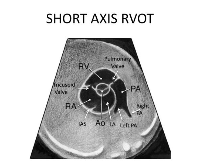
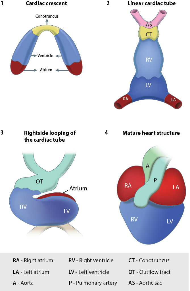
Cardiac week is one of the more difficult ones......which aspect(s) of the fetal heart are you struggling with the most? Respond to your classmates with helpful tips/tricks to remember these difficult concepts
Week 5: Forum/Discussion Board
Hi everyone!
I'll be the moderator for this board this week and I look forward to hearing all of the tips and tricks everyone has that have been helpful for them! It'll be especially helpful this week to share videos and images to help with visualization, as I know the fetal heart views can be difficult to wrap our brains around.
To start us off, something I am struggling with in terms of the fetal heart is definitely embryology. I have a hard time visualizing the fetal heart when it isn't fully developed yet and I struggle with the embryological terms. I think it would be helpful if there was a video or diagram that went week by week and showed the different developmental changes in the heart. I'll be on the look out for something like this and if anyone finds something that is helpful for them please share!
I'm excited to conquer the fetal heart with you all!
Hey Brittany,
I am right there with you when it comes to embryology! I find when things are color coded it helps my brain process new concepts better compared to when they're just labeled. Something that I am thinking about doing is sculpting the fetal heart in clay or using pipe cleaners to show the looping and twisting process that the heart must go through before it's in its final structure. I have so much clay left over from 1st quarters project, so it could be a fun way to learn this material!
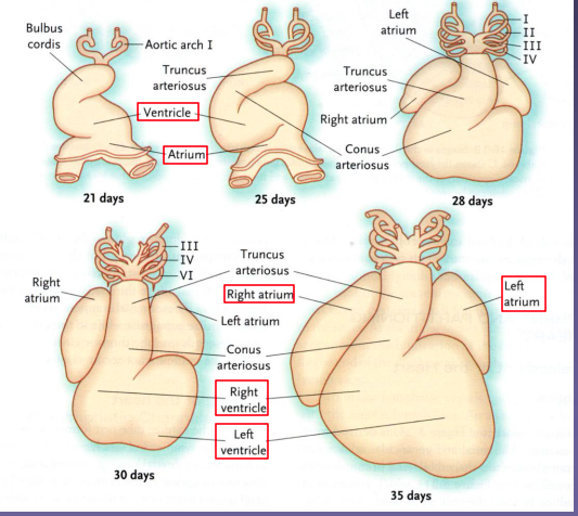
I also tried to find some images of the heart development week by week but I didn't find much information on the web! Here's another cool picture that is similar to the one in my original post that does include a timeline! Hope this helps!
Hi Chrishawna! Your suggestion of using clay sculpting prompted a youtube search that led me to this . She does a lovely claymation of cardiac development. I think I'm going to have to watch a million of these until it finally sinks in.
Leah and Alexis you get my vote for best video, that was SO FREAKING COOL!!! Everything I have seen so far has been individual pictures just describing the process but that was so cool to see it in motion! I think if I watch a couple hundred more times I might remember it lol! Thanks for sharing :)
Hi Chrishawna!
Thanks so much for the diagram it definitely helps! I think something I struggle with is how abstract the embryological structures look. I'm so used to the adult anatomy that my brain can't wrap around how it looks before it's fully formed!
Using clay is an awesome idea as well! Do you think you would create different models for the different stages of heart development? Would certain structures be color coded?
Hi Brittany,
Thats a great question! I think I want to start with it as one model made from different colors and try to manipulate it into different positions, but I’m not sure how successful that will be. If worse comes to worse I just might have to mold each step! The good thing is regardless of the way it’s constructed I should still learn something from it!
Hi Chrishawna!!
I have a feeling everyone is going to say they are struggling with embryology! especially because it just got introduced to us, it’s so new. hopefully one day we’ll look back and say oh that was easy! LOL. I love your clay idea. That really helped me when we all did it together so maybe we can have another clay date!!
Hey Chishawna,
The clay idea sounds amazing!! I definitely to do that too. I'd love to work on something like together if possible. I am unfortunately stuck at square one. I am not sure if I understand the loops and the twist to even begin crafting. Did you find any helpful videos to lock it in?
nailah
Hey Hailey & Nailah,
I definitely think we need to have another study group and have some fun with this topic! While it can be daunting I find that repetition is helping me better understand it. Sometimes I'll watch a video 4 or 5 times until I get to the point where I can explain the steps to others in a logical way. One video that was just released within the last week is by one of my favorite youtubers. He sometimes gives more info than we need for our purposes, but you can't deny that's he thorough! Heres the video if you want to check it out: https://www.youtube.com/watch?v=RjiPx6Xi-68
Yes repetition is key with this, I think i'll get it down a lot better after these 2 midterms coming up. It's so hard to retain everything at once. I know we are all doing are best and that's all we can do! I saw Paris mention a pip cleaner video so I went on a hunt lol heres what I found,
https://www.youtube.com/watch?v=p2Q5kI8uXIs
Hi Brittany,
I hear ya on the embryology! I wrote in another comment, I'm going to have to watch animations on repeat until I get it. , starting at 2:08, was nice and slowly went through each step. While it is difficult now, I know it will help me understand all the pathology as we progress in our studies.
I feel like I have a basic understanding of 4 main heart views (4chamber, LVOT, RVOT, and 3VV), and how one angles superiorly to cycle through them. I think one of my concerns is finding and keeping a good view while scanning. It is something I hope to practice in clinicals this week. Everything gets more challenging when baby won't stay still.
Also remembering all the things to check when scanning the fetal heart... maybe we can write a song or something? Some way to easily run through the long list in my head when scanning. I'm so nervous I'm going to miss something, especially anomalies or pathology.
I'll probably think of more as the week goes on, but these would be the initial 3 that come to mind. Can't wait to see what resources people share!
Hi Brit! Embryology is definitely a foreign beast. I found this short video (start at 2:07) that has great animations. I found it particularly helpful for the development of the septum primum and second secundum, and what structures form the septa (endocardial cushion and such). I'll likely draw these steps later since I find that helps me make concepts stick a little better. It also dives a little into what defects can arise, which is a great reminder of why we need know these processes.
https://www.youtube.com/watch?v=-d2UfOePgZw
Edit: Whoopsie, it's the same video that Leah just posted >.< I guess that means it is definitely a good one!
Awesome video Alexis & Leah!
It's great with visualization. I'll have to make a document with all of these links so I can go back to them frequently!
Videos are a great resource, but sometimes I find myself going down a rabbit hole and watching videos that aren't totally relevant. You're both so good at finding Youtube videos, how do you find quality ones? Do you search for something really specific or do you just sift through and watch a bunch until you find a good one? Do you have a specific channel you follow?
Sometimes my searches can be hit or miss. I typically have to watch a few before I find one that I like. If I don't find a good video within the first page of results, I'll try another search and change some of the wording. Like cardiac development to fetal heart development or cardiac embryology. Lots of trial and error, but it's worth it when you find a good one!
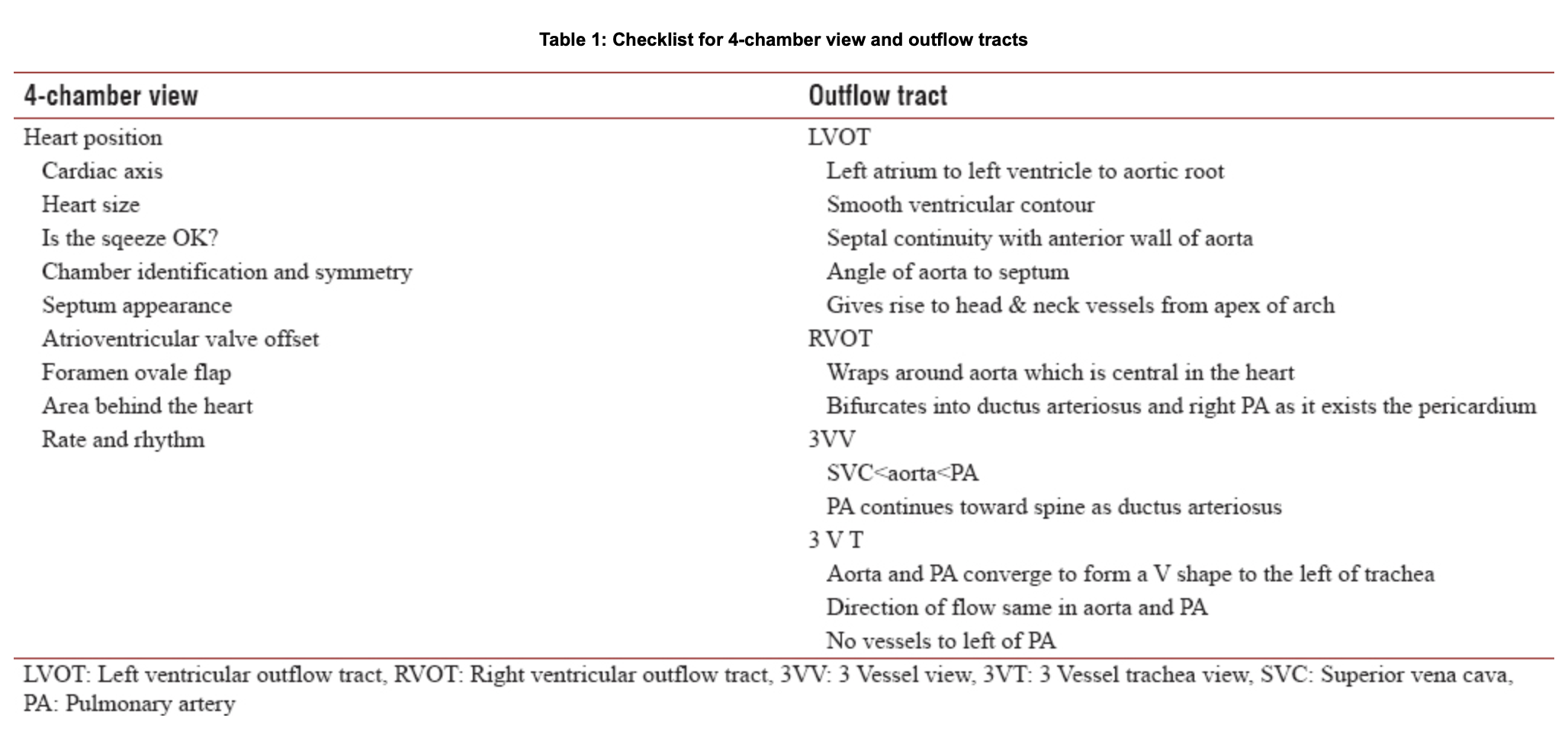
I think what I'm currently struggling with the most (besides the given, embryology) is how many things we need to consider and examine in each of the views. The lecture was slightly overwhelming and I was having a hard time organizing my thoughts afterwards. I just found this simple chart that lays out what we should be looking for. I think this is a great starting place for my own checklist and will definitely be adding on to it as I review the material more.
Hi Alexis!
I love the idea of writing out everything that you need to see in each view. I think it's a great organization tool.
How does your CI keep track of all the images that need to be taken?
I haven't seen an OB anatomy scan yet so I'm not sure! For other exams, my more seasoned CI knows all the protocols like the back of her hand and my other CI uses scan assist. I don't think I can program these types of cues into scan assist, so I'm sure I'll have to make my own checklist and keep it in reach when I scan in the future.
I am in the same boat Alexis! But I compiled a small pop quiz for you to differentiate views. I will grade easy don't worry. What view is this?

There is another lecture that is up, I'm not sure if you guys looked at it, but when I was a student it helped me A TON! Everything is broken down into nice little sections and the images are incredibly helpful! It's at the bottom of the week 5 class shell and it's called Normal Fetal Circulation. I rewrote this ppt in my notes and drew the pictures that I found especially helpful!
Other than that, this is a heavy topic. EVERYONE struggles when learning it. Even Michelle did when she was a student. Just keep on keepin on :)
Cardiac week has definitely been the most difficult thus far. With that being said, there are some concepts I feel I have a good grasp on and others that I am struggling with. In terms of the overall fetal heart anatomy I think I have a good understanding of the anatomical structures and how they connect. I have been working on drawing out the process of fetal circulation and am feeling a lot better on that aspect as well. I have found that if I can draw it, I can understand it. The area where I am struggling the most is embryology, it's such a complex and delicate process that it's easy to miss an essential step. I am hoping to spend this week binging videos on this topic and hopefully drawing it out from the early stages of the heart tube to its final product. Sometimes recording myself saying the lecture material and playing it back helps me retain as well, I am also going to try that this week and see if I am successful!
https://teachmeanatomy.info/the-basics/embryology/cardiovascular-system/
Also I love this graphic because each part is color coded making it easier to follow!
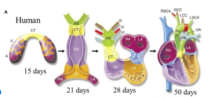
Hi Chrishawna,
Thank you for sharing that you record yourself going over lecture material! That's definitely one way of studying I've never thought of.
I, and the rest of the girls, I'm sure, would love to see your drawings!! Your notes and drawings are always so organized, clean, and helpful.
Love the diagram you shared, too!
Thanks,
Karen
Karen you are too sweet! You know me, I have to doodle to get it in my noodle lol! Some of the other girls mentioned this as well and I think it's super helpful to review the adult heart before conquering the fetal. If you know the basic anatomy you just have to remember what the adaptations are! Since you asked, here's a quick sketch I did a few days ago to help me!
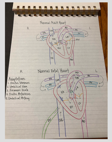
Chrishawna,
Drawing it can really help! Remember to use different colors to help the structures pop out.
I found this video extremely helpful. Fun fact: it's super old but still useful. https://www.youtube.com/watch?v=5DIUk9IXUaI
Sarah
Like the rest of the girls how have already posted, I can tell embryology is going to be a hard one for me. I was really trying to understand everything Dr. Wilson was lecturing about but it was really difficult. Like she said, we really need to conceptualize this topic and visualize it in our minds and that is gong to take some time for me.
I found this video that shows the process very clearly.
https://www.youtube.com/watch?v=a0qyagIgBPw
Hi Hailey,
I'm totally with you on having a hard time understanding embryology!
What do you think is the most daunting part about embryology for you? Do you struggle with the vocabulary or visualizing the developmental steps?
Hi Brit!
I feel like vocab might be easier to memorize compared to the visualization part. It's like memorization vs really understanding. The understanding and visualization is going to take alot of time for me to really get. I'm really glad that understanding is how I learn though because I feel if i understand something vs just memorizing, I can really think things through which is so important for OB. It'll be be more difficult, but also more worth it in the end. what about you?
I'm with you on the visualization!
I'm so used to fully formed adult anatomy that embryological structures seem so abstract and foreign. They almost feel "wrong" because they don't look like how they should! Well, they don't yet anyways!
Check out Chrishawna and Karen's posts. They have great images of the embryological stages of the heart!
Hey Brittany, that's a great though! Sometimes it's helpful to build off what you already know. Maybe it would be helpful to look at some adult heat images to get a grasp on the "big" picture. Then think about how those areas came to be.
Hi Brit,
Totally agree with you on the feelings wrong part! It’s so different to us so it definitely is going to take a while to get used to. I can’t wait to put all my energy into this after our midterms, I want to understand it so bad! LOL I guess it comes with time.
I will check their posts thanks so much!
Hi everyone!
I definitely think being in clinical has helped me with identifying the heart structures. However, I'm having a hard to conceptualizing and envisioning where I should place the transducer on the mom's body in order to get the views I need. I want to be able to intentionally place my transducer down and know what angle I'm going to get instead of luckily just stumbling across the view I need.
I found this image and it seems to help me a bit in my head to visualize it, I'm going to try to apply it tomorrow in clinical and see if it helps.
Here is the image that goes along with Lania's post! Her computer was having some technical difficulties :)
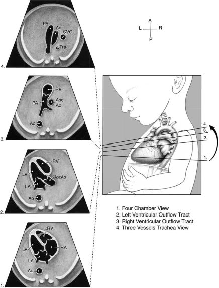
Yes I love this!
The next time you guys are at lab, as Sue for the heart model. It's a full adult heart that breaks up into scanning planes. That heart helped me to really conceptualize the way to angle my probe to achieve the different images we need, just like this image! For those of you that like hands on, you'll hopefully find it beneficial.
This image helped me conceptualize each of the views and features. Thank you for sharing girls. Narges told me the three vessel view helps rule out 90% of cardiac abnormalities which makes it a super important image to capture. Does anyone else have tips to scan these views bc I loose the heart when I move cephalic.
Howdy Lania,
Clinical helps a lot! I'd love to have a few 20 week moms in lab. There's nothing like hands on practice.
Sadly there really is no "correct" place to put the transducer on mom's body (in my experience). It's totally dependent on how baby is positioned.
This image only shows a cephalic presentation BUT baby can be in so many positions! So the transducer doesn't really have a home.
Sarah

H Sarah!
You are absolutely right! I learned today that honestly the position and views can be attained from anywhere, which actually gives me anxiety lol mainly because I like structure and knowing an organ or structure should be in the same vicinity each time I put my transducer down. So, this is a bit of an adjustment for me honesty. But as you mentioned, there's nothing like hands on practice to make us better and feel more confident.
Hi Lania,
I completely agree with you! OB is nerve wracking because the baby's anatomy could be anywhere within the uterus at any given moment. It kind of reminds me of the spleen. Depending on the patient sometimes its easy to get those images no problem and other times were rolling the patient all around just to get a glimpse of what we need. I want to work more on understanding fetal lie, if we can get that down I think it will be really beneficial!
Hey all,
Honestly I have learned the heart many times before and it has always been a foreign concept to me. I think because of how intricate and complex it is compared to the other organs. When you add on embryology and shunts it is a whole other ballgame. However after this past week I have learned a lot about the fetal heart and am somewhat starting to get the hang of it. Something that I thought I understood but realized I didn't was the septum primum, septum secundum and the foramen ovale. I found this video that helped out a bit.
https://embryology.med.unsw.edu.au/embryology/images/5/53/Heart_septation_003.mp4
I am also struggling a bit with the RVOT. I am confused on the branches of the pulmonary artery. I know we have the RPA which courses toward the right and the LPA which courses to the left. But after looking up multiple images I find that the LPA is labeled LPA or ductus ateriosis. I know that the ductus arteriosis connects to the LPA but can we actually see this? They look the same to me but I see them labeled on or the other. Any help is much appreciated!
Labeled Ductus
Labeled LPA

Hi Allison!
That is confusing how some images label it LPA and some label it the ductus arteriosus. The only thing I can think of is perhaps they're in a certain plane where they only see one or the other? They both branch off of the main pulmonary artery close to each other.
Also, the ductus arteriosus can be seen entering the aorta in what is known as the "hockey stick sign".
Can you find any images of the "hockey stick sign" vs the "candy cane sign"? What different structures are seen?
Hey Brittany,
So I found this. This would be the hockey sign. It does look like in the sagittal view that the ductus and LPA kind of bifurcate so maybe in the RVOT view the ductus would be more cephalad? I honestly don't know.
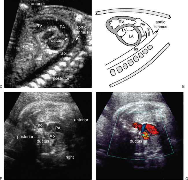

Image F on this last image I think shows it well the ductus is a bit superior to the LPA. So I would assume now in the RVOT view if you are more superior it is the ductus and more inferior then the LPA.
Also image E is the "Candy Cane".
Awesome images Allison!!
I really love image F, I think it shows the ductus arteriosus connecting to the aorta really well. Images E and F are also really great ones to be able to identify and differentiate. The "candy cane" image shows the aorta and is a bit rounder than the "hockey stick sign" showing the ductal arch.
Great job looking into this further!!
Allison, I love that you brought this up! I have asked SO many different techs about this to try to understand better so here are my thoughts:
They're basically both correct. It's hard to explain without visually showing you, but basically, in this view there are structures we can't see because of how our probe is hitting these structures. We can see the RPA branch as it heads to the rt lobe of the lungs, but the LPA is actually shooting backwards, parallel to our sound beam (remember the heart lies slightly at an angle with the RV more anterior and the LV more posterior). I did my own labeling on this image so I hope it helps.
You can see the split between the RPA and LPA before the LPA dives and then we are see the ductus arteriosis (DA). It originates from the LPA and dumps into the descending Ao (DAo).
Hopefully this didn't confuse you more!
Thank you for the explanation, Paris! I looked up an adult heart CT video to see what you mean about the LPA and RPA branching in different directions. The still below really helped me visualize what you were saying about the LPA shooting backward and why we aren't seeing a larger chunk of it in the RVOT view. 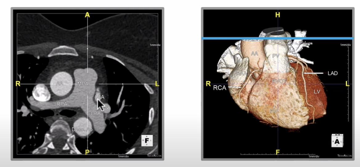
https://www.youtube.com/watch?v=NSHm-fl5Vsg
Paris!!
Thank you so much. No it did not confuse me. It makes sense! Essentially sonographically it looks like one vessel however since the LPA dives backward we would only see a small longitudinal portion of it. Then at the same level we see the ductus. Add the two together and it looks like one vessel. Am I somewhat on track here with my thought process?
What makes this week even more difficult is that it's also the start of our midterms. So I'm just rolling with the punches. As we begin to dive deeper into the fetal anatomy, the heart is weighing heavy on me. It's hard for me to lock things down because there's so many different pathways or the fact that so many different anomalies can begin and it just throws off the whole development. It feels super overwhelming, but I really just need to break everything down. So understanding few of the basic concepts/ structures of the heart is something that I NEED to solidify before begin to continue. I need to build a strong understanding and foundation of the fetal heart, so that everything else will come...just a bit easier. Throughout the week, I plan on diving into the embryology first. Chrishawana had already recommended to use Khan Academy and to draw everything out!
Hi Monica,
I definitely agree that this is an overwhelming week, but I think it's normal to feel this way! Something that helped me with fetal circulation is first understanding normal adult heart anatomy and function. Once you have this down I think it's easier to go back and look at the different parts the fetal circulation bypasses.
What do you think would help you most with learning this material? Drawing, listening, watching videos, reading?
I agree with you Brittany! I realized I was having trouble understanding the video lecture since it’s been a while that I’ve thought about the heart. I watched this short video to help refresh my memory about circulation through the adult heart. It helped give me some context.
Hola Brittney!
So I plan on solidifying my understanding in knowing the fetal heart structures and drawing everything out, then build up from there.
I talked to my CI about how I should be planning my study material out and he agreed with my strategy. But he also reminded me that everything will click next quarter! So I'm holding on to that hope lol
Hi Monica!
You are doing great, hang in there, I think we are all feeling like this is a lot of material but remember to be patient with yourself! You will get it. I found this short quiz really helpful in making sure I understood the basics of fetal heart anatomy and circulation, it might be helpful to you also! I also like viewing multiple videos of the same thing, because hearing it from different voices helps me remember it!
https://www.registerednursern.com/fetal-circulation-quiz-maternity-nursing-nclex/
Hi Monica!
I completely agree with you! I think we're all feeling super overwhelmed right now, but the good thing about these boards is that we can bounce concepts and knowledge off of each other. Someone's weakness may be someone else's strong suit! I think the best thing we can do right now na try to scan the fetal heart as much as we can in order to lock these structures in. I know I'm personally a visual learner, textbooks do absolutely nothing for me lol so seeing it in clinicals and also I've found drawing each view out has helped me a little bit more lock it in as well. Maybe that can help you as well? :)
Hi Monica,
It seems we are all struggling, huh? I definitely feel the same. However, you are so great at creating study materials, I am not at all worried about how you will handle all this new info. Remember all the great slides you made for physics when it was overwhelming?? Not to mention all the flashcards you made, don't forget about those!
Do any of those anatomy apps you have help with teaching the structure of the heart? I haven't even had any time to check for any!
You'll do excellent in this class.
Karen
Hi Class,
I have a long list of concepts I am struggling with this week. However regarding the fetal heart, the fetal heart views is something I am having trouble conceptualizing. Specifically what I am supposed to see in the LVOT, RVOT, three vessel layer. I know I should see 5 different compartments in LVOT but I am having trouble remembering what those 5 compartments are.
I am also having trouble remembering where the blood travels to in the fetal heart and how that differs from an adult heart.
Today’s second half of lecture about where the different parts of the heart form from was really difficult for me.
I have watched multiple YouTube videos but still having a hard time since there’s so much to remember. However, I would still love any extra videos you guys find.
Hi Candee!
I agree that the outflow tracts seem super tricky along with embryology! It helped me to memorize the order that we take these images in to understand where I need to be to obtain them.
For example, we typically start our cardiac images at the four chamber heart. We know by sweeping more cephalic we should next hit the LVOT, from there sweeping more superiorly we will encounter the RVOT and finally we should see the 3 vessel view scanning up even further. So if we're scanning the heart and we suddenly go from the 4 chamber up to the RVOT we could assume that we might have swept too fast and should come down slightly inferior and see the LVOT in between those two views. Again this is assuming that the baby has a normal anatomy and that the outflow tracts do cross.
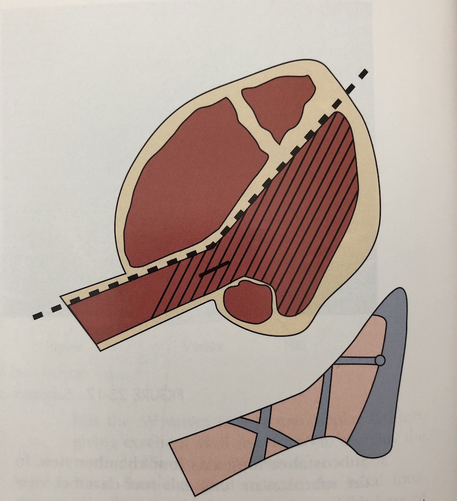
One image that I always keep in the back of my mind for the LVOT view is a ballet slipper. I attached a picture down below so you can see exactly what I'm talking about, but if you see the ballet slipper you know you're in the correct view! Hopefully this helps!
Hi Candee,
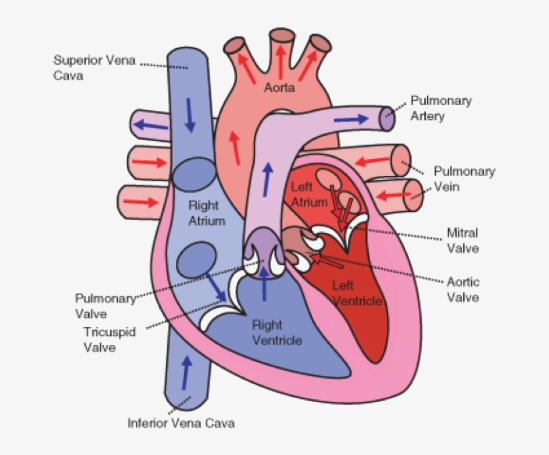
It can definitely be hard to keep track of the blood flow path in the heart. I think it's beneficial to go back to the basics first and review the normal adult heart. I've attached an image here with arrows to show flow in an adult heart.
I think it helps to remember that the adult heart basically works as two halves. The right side pumps blood to the lungs to get oxygen, and the left side pumps blood that returns from the lungs out to the rest of the body. Once this becomes a bit clearer I think it's easier to then go back and add in the bypasses that the fetal heart uses. It helps me to remember that the fetus really wants that oxygenated blood fast so it doesn't want to take long routes and instead tries to get into the aorta asap!
Is there a specific part about fetal circulation that is challenging for you? Is it just difficult because the fetal heart has several different routes?
I think it's just remembering the different parts that is difficult for me. But the pictures are definitely helping out.
Hey Candee,
I get what you saying. You know the heart its just not second nature yet to spill it all out. Things that really help me out are
- ATRIUM = an open-roofed entrance hall. Think of these as the entrances. (IVC, SVC, Pulmonary veins)
- The LEFT ATRIUM is best friends with the AO they are always close.
- The RIGHT VENTRICLE is in a BAND the MODERATORS
I always think of these 3 things that help me start to figure out where I am.
Like a lot of us ladies, I think what I struggle with the most is embryology and formation of fetal heart. Not to mention circulation, which I'm sure if we fully understood and memorized, would help us immensely on the different segments of the heart that we need to image!
I found a great short video that walks us through the development of the heart:
https://www.youtube.com/watch?v=a0qyagIgBPw&ab_channel=HyeonjooKim
Today's lecture was a little overwhelming, to say the least. But, I think this week's assignment of writing a "story" about circulation will really help our understanding of this very important topic. I love and am so grateful that our group is so sharing with helpful videos and tips, in these discussion boards and in our group chat.
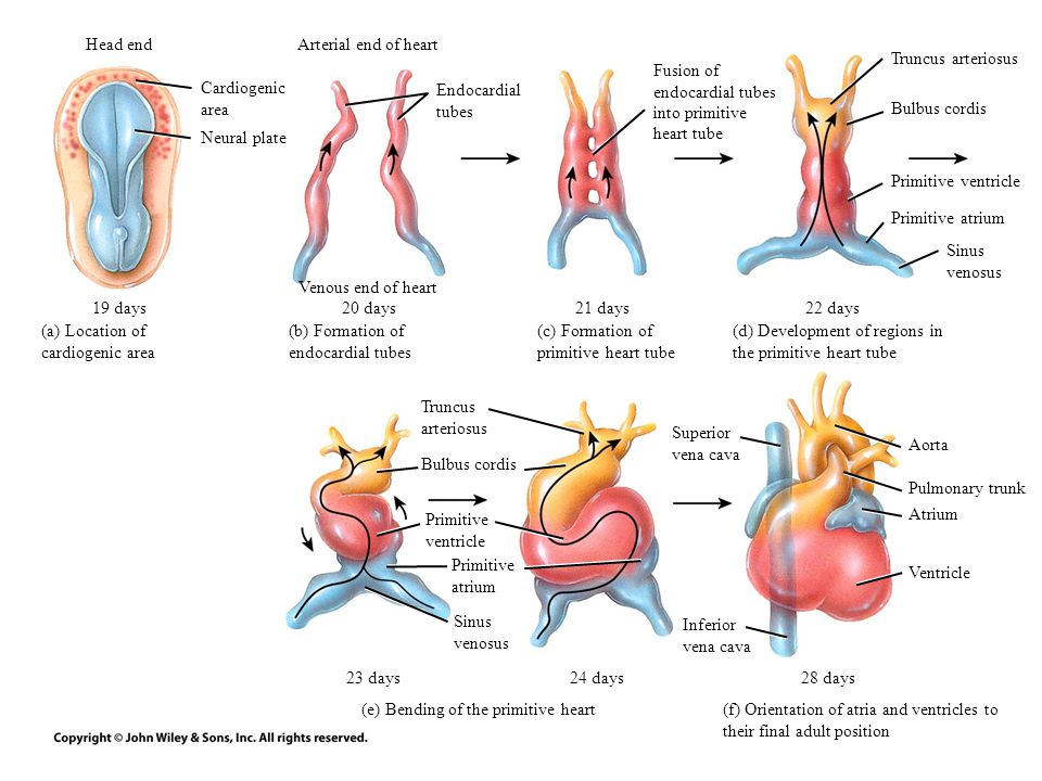
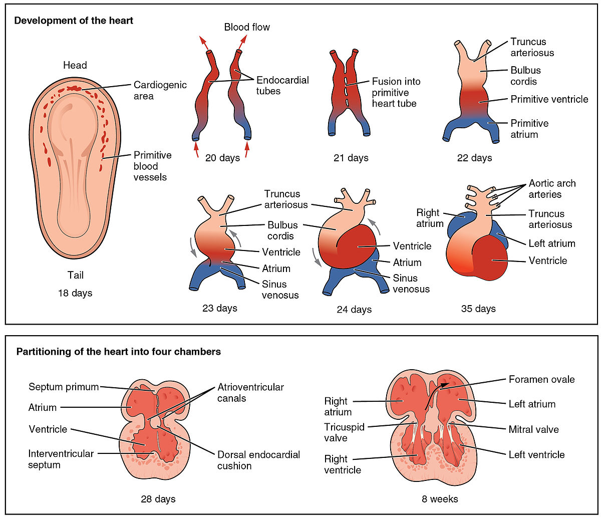
Here's a diagram of formation of the primitive heart:
Hi Karen!
fetal circulation is another beast we have to get down on top of embryology. It’s going to be rough but I think the stories we create are really going to help us remember. I found this video on fetal circulation, I always love watching this guys videos. He’s really good at explaining and drawing everything out.
Hi Karen!
I love this image you shared. Embryology is a challenge for me too and I think it helps to see images of what we are starting with and where we want to end up.
What do you think is the most challenging part of embryology of the fetal heart for you?
Hi Hailey,
Thanks for the video! I will take any extra help I can, haha!
Hi Brittany,
To be honest I think it's really the extra terminology and remembering what becomes what... I feel what is partially overwhelming is all the information we have to digest and memorize this quarter. My brain can't keep up with the constant waves of new information!
All in all the "repetitiveness" that Dr Wilson is going for should help us start to fully absorb everything.. or at least recognize, which is a great start. Still, it is hard not to feel like we're almost drowning in this quarter's classes.
I'm glad we're such a great and supportive group.
Thanks ladies,
Karen
Hi everyone!
This week's topic on the fetal heart has been more so challenging for me than prior weeks topics for sure, but it is making me work even harder to understand everything better. I really wanted to simplify the heart in my presentation, but it seemed like there was information too important not to mention. It is definitely an ample amount of information to digest. The part that is giving me a hard time is for sure embryology and also trying to obtain the RVOT, LVOT, and 3 vessel heart views on the biometric at school. I haven't gotten much back scanning time in at clinicals with OB yet, I've only taken a few heart rates at the 4 chamber view and some cine clips needed for a survey. I need to work on orienting myself with the baby's lie. It seems like it should be so simple, but when the baby is moving all over the place it seems like it would be challenging to maintain awareness of the fetal lie while imaging those important landmarks. So I think it'll definitely take some practice with my CI to obtain the different views. Any tips from would be much very appreciated!
I found these videos helpful:
Hi Molly!
Great videos, I actually watched these before class on Monday! They definitely help me visualize where I'm scanning on mom as well as what image I'm getting of the baby.
I struggle with determining the baby's lie as well. I did notice last week though that our biometry simulator lets you alter the fetal position! It may be helpful to practice getting all the measurements on one setting and then changing the position and ensuring you can still do it!
What do you think would help you with visualizing the position? Maybe using a doll? Drawing out the views for each position?
It also may help to start with looking at a specific landmark each time. I know for me I first look at the spine and then try to orient myself!
Hi Britt!
Great job with Moderating this week! Wootwoot! That's awesome you watched the videos as well! So great to find resources like these online!
Thank you so much for sharing that the biometric has the ability to let you change the fetal position. I did not think we were able to do that! So awesome!!
I think using a doll is a super smart idea! I'll also try with drawing out the positions, I think both would help a lot!
That is super smart of you to look at the spine first to orient yourself! I see the Sonographer's start with the spine when baby is on its stomach. When they're on their back they usually start with the heart.
Thank you for your help!
-Molly
Hi Molly,
I love these videos! It made a huge difference for my understanding of the fetal heart views. I'm also grappling with the concept of fetal lie. I was just talking to one of the CIs at my site about how there is my Rt/Lt, the screen's Rt/Lt, mom's Rt/Lt, and baby's Rt/Lt. And when you consider baby can be upside down and facing any which direction, it is definitely an art that I'm trying to master. I've been trying to watch the beginning of the scan when my CI does it and then determine fetal lie. Today I got one right!
Keep up the good work!
Hey Molly,
I am with ya girl!! The heart is hard!!!! I think my first priority is trying to really nail down fetal lie and where the heart should be. I found this really interesting image that shows if you assess the fetal lie properly this is where the heart should be. If you close your fingers and extend your thumb the babies head signifies your knuckles, your palm side is the belly, back of your hand is the babies back. If you extend your thumb it should signify where the heart is. It looks interesting, not sure if it actually will help but its a start!
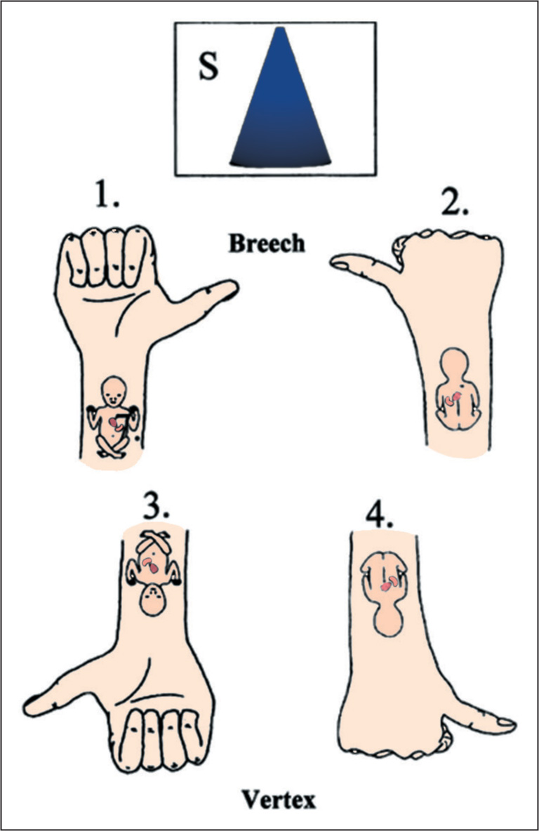
Great videos!
Try to see if you can find one for RVOT in an axial view of the heart. For LVOT we like to see it in a long view, but for RVOT, we like to see the split of the pulmonary arteries in addition to where it leaves the RV. In order to see both, we have to be in an axial plane.
Hi Molly! I definitely had some false confidence going into this week about fetal lie. It all makes sense in my head, but you're totally right that the baby moves around and makes it so much more difficult! I have gotten one thing drilled into my head these first two weeks in clinical though, and its to do a sweep before imaging anything! My CI tells me often "no, sweep through first" when she sees I'm jumping into that first image. I think doing that initial sweep is super helpful for determining fetal lie since we will go from possibly abdomen/chest area (wherever you put your probe down) and sweep around to find possible head or feet and then be able to sweep through the whole length of the body again to see which way baby is lying. I hope this little tip helps!
Hi Lauren!
That tip definitely helps! Our first images are to always do a cine clip of the baby in long and in transverse, I like that it forces me to orient myself. Although it is tricky! Also especially tricky when viewing the baby in transverse especially the spine. The tech will point out the C,T,L & sacrum spine in trans and I feel a bit confused. I can recognize those parts of the spine in longitudinal but transverse throws me off for sure. Luckily my CI wants to take OB slow and focus on one part of the body at a time. Currently we‘re working on me just doing a cine clip in both planes, getting moms adnexas, measuring baby’s heart rate, and beginning to scan the head and get proper measurements. Today the baby was 19 weeks along and we had a hard time trying to get the CSP show that perfect square box in order to get our head measurements. My CI emphasized the importance of that CSP showing nothing within it and for it to be a nice clear image. I had to keep making adjustments with the probe and my CI helped. Highly recommend focusing on one part of the baby and working up to more over time.
Brittany mentioned that the biometric allows us to change the fetal position! Can’t wait to check that out to help practice determining fetal lie and getting those landmarks.
Howdy everyone,
Man, it's hard to choose just one. I think my main confusion right now is how exactly to switch from LVOT to RVOT and vice versa. Sweep superior but also rotate clockwise? I'm need to find some videos on this.
Also, I know 4 chamber should be easy to get but I'm having trouble getting all 4 chambers open.
Also embryology - but that one should come with a few more reviews!
Sarah
Ello Sarah!
I'm totally sitting in the same boat as you!! I'm excited to go to class on Friday and practice with the Mommy phantom! Dr. Wilson mentioned that it's a great resource to use and help us lock in the technique. However, nothing is ever like real life... on a baby that moves around every other second! Lol
All I know is that it's a huge struggle right now, but my saying is Just Youtube It
With embryology, we should all draw it out together or do another clay project. It's a lot to digest, but we will push through!!!
Cardiac week is definitely challenging! I think some aspects of it I am finding difficult include embryology, understanding how everything forms and develops. Scanning the fetal heart in clinic is also a bit difficult, although I have obtained heart rate with M-mode. Even though I can get the heart rate, I am having a difficult time obtaining the 4 chamber view, and being able to bring the heart into better view so it's not obstructed by shadows. After I determine the fetal presentation, I try to start at the head, and scan inferiorly on the fetus to land on the heart, but I'm finding it challenging to do so. I think this will take practice and I'm sure we will all master it, but if anyone has any tips I'd love to hear!
I found this fun short video "10 second quiz" to see how we we are coming along in being able to identify "abnormal" in the fetal heart!
Hey Zuly,
I am really struggling with the embryology too. I typically use videos to lock everything in and I have to find a good one. I am going to check out the series you posted to see if it helps. I also can empathize with obtaining the four chamber view struggle. It is not easy because the angle of insonation can pick up extra anatomy. Only tip I have so far is keep moving around.
Nailah
Hi Zuly!
I can only imagine how hard it is to get these fetal views in real life. Have you been able to ask your CI what helps them? Or observe what transducer movements they do in order to get around shadows? It also may just be something that takes practice and eventually becomes muscle memory.
I remember when we all started scanning the liver and we were struggling to find landmarks like the left portal vein, and now look at us! We will get there with the fetal heart too it just takes time.
Hi Brittany!
So far I haven't seen a study where the techs at my site obtain the 4 chamber view, maybe I just haven't gotten lucky yet! We do get a lot of AFI exams so maybe I just need to wait until I get a full anatomy scan to be able to pick up some tips! I'm sure it will come with practice though as you said!
I am having a hard time understanding the LVOT and RVOT both anatomically and how they work into fetal circulation. I think that's because most of my understanding of the heart is coming from my basic anatomy class and we didn't discuss those 2 (or I don't remember it lol).
I was also struggling with visualizing the ductus venosus and ductus arteriosus, but I found this hand drawn image that helped me! I understand that the 3 shunts are as a "shortcut" for blood to get out to the body, but I wasn't picturing them well!

Hi Lauren!
Great visual. From my understanding, the RVOT and LVOT are basically just the pathways of the pulmonary artery leaving the right ventricle (RVOT) and the aorta leaving the left ventricle (LVOT).
It really helped me to remember the basic functions of the adult heart and to recall that the right side works as its own system pumping to the lungs, and the left side works as its own system pumping to the rest of the body! From there I think it's easier to then think about the different bypass routes that the fetal heart takes.
When scanning, are we able to see the 3 fetal shunts? If so what should they look like? (ductus venosus, ductus arteriosus, and foramen ovale)
Hi Lauren!
I love this visual! It definitely helps show the flow in an organized way. I was also having a hard time figuring out how the shunts played a role in the circulatory system. Along with this image, the diagram Paris made has also been super helpful with showing exactly how everything flows and what the different paths back to the placenta are.
I liked this explanation from Stanford children's health too. It is super straight forward and goes through each step of circulation.
Blood Circulation in the Fetus and Newborn (stanfordchildrens.org)
-Molly
Molly, I read your linked article and it really helped me understand the fetal heart circulation better! I'm glad it touched on the foramen ovale and ductus arteriosis closing at the end because I was curious about that; I remembered foramen ovale being on the heart model we studied in my prerequisite anatomy class, but I wasn't sure how/when it closed off. Thank you for the great resource!
For cardiac week I think I am struggling with the big picture. I feel like I have bits and pieces but having a hard to time understanding how they all fit together. In terms of cardiac embryology, to my understanding:
- Cardiogenic cords
- Cords develop into a single endocardial tube
- The cephalic portion of the tube bends right and forward and the caudal portion bends left, backward, and towards the head simultaneously.
- These morphological changes form an atrioventricular loop. I am not sure if this bulbus cordis?
- This loop develops into the atria and ventricles. How? I am not really sure. Maybe the twisting of the loops develops these structures
- Endocardial cushion separates the atria and ventricles.
- These cushion grow inward, developing the mitral and tricuspid valves.
- Somehow the ventricular septum is developing at this time as well but I am not quite sure how. However the muscular portion develops first then the membranous
- Septum primum seprates right and left atrium. I am not sure how the structure arose, maybe from the membranous septum?
- Septum secundum is formed (not sure how)
- The aorta and pulmonary artery arise from the truncus arteriosus, which essentially twist creating these structures.
- The development of the aortic arches confuses because I am not sure how this process works fully. To my understanding six sets are involved somehow.
I am missing quite a few pieces. I am struggling finding a good video as well. Any visual examples would be great. Thanks guys!
This image is a nice visual:
Hi Nailah!
Wow look at you already being able to list out embryological steps!! That's awesome and it definitely helps to list out what you know and what areas you need clarification on.
I also love this graphic. What resources are you finding most helpful for you? The textbook, our class lectures, Youtube?
Hey Brittney,
Thank girls. I feel like I am STRUGGLING though for sure. I'm missing pieces : ( . I found the majority of this information in the book, but I live for YouTube videos. I haven't found any good ones yet. Chrishawna suggested this one:
https://www.youtube.com/watch?v=RjiPx6Xi-68
I have to check it out.
Hey Nailah!
Great images and explanation! I'm going to need you to help me with the embryological cardiac development it looks like lol. But as I was studying today, I realized how important it is to understand the embryology aspect first in order to understand what could possibly have went wrong if we happen to sonographically find a heart defect in the fetus.
Hello everyone,
I feel like I am struggling with everything at this point :( In terms of the fetal heart, I am understanding the different flows and pathways but I am having issues with scanning it. I can get the 4 chamber heart but when having to identify the RVOT and LVOT I am not getting it. I was scanning a baby today actually and I got images but just could not differentiate them that well. I think I need to study more on following the actual ventricles and studying more pictures to hopefully get a better understanding. Any suggestions?
Here is a video I found that is helping me a bit: https://www.youtube.com/watch?v=YTmx3e24erc
Hi Raman!
You and I both girly! I'm completely the same way so don't feel alone on that! I think we have to be kind to ourselves and realize that we are still in SCHOOL for a reason lol. We will get there but we have to be patient with our learning process. It can be hard when we're in such an accelerated program, but as long as we do our part and study as much as we can, IT WILL eventually make sense. I've been watching YouTube videos like crazy to help me lock everything in!
Hi Raman!
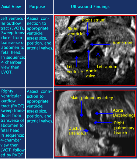
I'm sure it's quite an adjustment to go from reading about these heart views to actually scanning them. The biometric simulator at school is actually pretty helpful for getting used to scanning though! It shows the outflow tracts and you can change the fetal position to get used to looking at the heart in different views. I've attached some images that are labeled and will hopefully help too!
Does your CI have any tips or tricks that help them to recognize the outflow tracts? Maybe associating the views with other objects would be helpful, I think it was Chrishawna who found an image of a ballerina slipper that looks like the LVOT!
Hey Raman!
I'm 100% with you. It's a lot to learn in such little time. We should continue to draw out the images of a fetal complete protocol and then list out what we should see. I find the the other discussion post this week, really helped me retain so much info about the 4 chambers. I hope that advice helps you out too.
I'm also finding it hard to image RVOT and LVOT as well. When I asked my CI for advice, he was telling me that it will all come with time and experience. Practice, practice, practice and it shall come to us in second nature.
Let's keep pushing through this week!
WE GOT THIS!
Hi Raman,
The fetal heart is so humbling! I've seen so many videos of OB scans and the sonographers make it look effortless. One thing I always say to myself when I feel that I am struggling is "look how far I've come". Six months ago we knew nothing and now we have tackled abdomen, are working through GYN, and doing everything we can to grasp OB. I think our mentality plays a huge role in our abilities, if we believe we can, then we can! It sounds like your doing great in clinicals, and the more you see it the easier it will get!
I totally understand that feeling, Raman. It's so frustrating too when you're scanning a patient and you can tell you're so close to the good image, and your CI comes behind and BARELY moves the transducer and its a beautiful image. I hope once we get the textbook of this down, the bits we get to scan in clinicals will lock it in for us instead of discourage us!
Totally, Lauren. It's all about the micromovements! It is definitely a skill we're all working toward and we'll get there with more scanning time.
I just rewatched this video I came across in the first quarter about planning your movement to optimize your view. Sometimes its good to refresh the memory and go back to basics!
https://www.youtube.com/watch?v=OHl-fxefyH4&t=11s
Great job everyone!! Now that we have discussed our concerns and worries a bit I wanted to pose another question to you all to talk about how to tackle those fears and feelings of stress.
I know I cannot study well when I am tired or feeling frustrated and I often need to take small breaks to clear my mind before returning to studying. For me, this often entails taking a little walk outside and visiting my horse! I also like to break up studying into small chunks so I don't get burnt out.
How do you all deal with feeling stressed and overwhelmed when learning a new topic? What helps you to not get frustrated and study your best?
Hey Brit!
I actually and still trying to learn what works best for me when I need to study but I'm feeling stressed and overwhelmed, especially since it seems like I'm always feeling that way lol. But so far, like you taking small consistent breaks while I'm studying I've found helps me stay focused a bit more, however I've found it 10x harder to find the energy after clinicals to come home and study. So I'm trying to figure out the best routine for that as well.
Oooooo I see you moderator!
When I start feeling overwhelmed I take a step back and list out my game plan. I typically take an outline or the objectives of the textbook and answer all of them out.
What helps my frustration and relaxing my mind, so I could study the best is working out. But it's so hard to fit that into this schedule right now )-:
Great question Brittany!
It's definitely very difficult finding time and energy to study now that we have clinic. I sometimes just try to remind myself to take things one week at a time, and honestly lately, just one day or even hour at a time! It's all so much but I try to just focus on one thing at a time, watch a YouTube video explaining what we're learning, drawing things out, making small outlines, and taking very short practice quizzes. Just one task at a time!
For me, I find it easiest if I plan out my week's studying ahead of time and pace myself so I know exactly what I need to do and what day I plan to work on it! I have also found that making super simple outlines of the chapters (basically just organizing the headers and subcategories) really helps me keep everything straight since it can get confusing where you're at in such dense chapters! I really appreciate our group always giving each other our study methods and sending links we find helpful, since if it helped one of us, it is bound to help someone else with similar learning styles.
Hi Brittany,
Mental health is so important and as students it is often put on the back burner. If I'm feeling stressed or overwhelmed there are a few things I always do:
1. Confide in my peers (who knows the struggle more than those experiencing it with you)
2. Walk it off- Literally and figuratively (might take Bentley on a walk or move onto another subject for a while)
3. Take some time for myself (This has been hard with everything going on but I might take 10 minutes to give myself a facial, paint my toes, or take a bubble bath)
Stress is unavoidable in life, its more about how we manage it!
You all have such good responses! I know it's easy to feel like you're drowning but it's important to take things one day at a time and break studying up into small sessions.
Chrishawna, I love what you said about self care!! It is incredibly important to take care of ourselves during this time. A lot of you mentioned just taking a small break or looking away from the topic for a while and that's a great method. We also need to remember to give ourselves the basics of care such as eating good food and trying to get a good amount of sleep. How can we expect our brains to absorb everything if we don't nourish it and allow it to rest!
Also, I had a chemistry professor tell me once that sleep is so so important when you're learning new things because it allows your neurons to make connections so you can process the new information and remember it!
I know there aren't enough hours in the day but try to take a little time to care for yourselves!
Hello Brittany,
I always try my best to not get overwhelmed and to combat it I work on my inner dialogue. So for example, let's say I had a rough day at clinics, instead of beating myself up for it I reassure myself that it happens to all of us, even our CIs. Plus, comparing ourselves who are barely a month into training is very silly if you think about the fact that some of the techs have over 21 years experience. One tech that I know has been doing ultrasound for longer than I have been alive haha. I am very lucky to have a nice support system and one thing that I have been doing is if something bad happens like I get a bad grade on the quiz (like this past Monday's) than I only talk about it once like to my boyfriend maybe and then move on. Also, breaking down everything per week, starting with the most important assignments first and knowing that realistically to actually learn you should not need to be studying all the time but rather need sleep and taking breaks is key. This may sound cheesy but you drown not by falling into a river but by staying submerged in it. So stay positive ladies. :)
Hey Allison, Candee, and Leah!
Allison, I really love the images you shared with helping determine fetal lie and situs! I remember this technique being mentioned during class, but wasn't sure if it would be a good technique. I will definitely try it out! Thank you so much for sharing!!
Candee, The variation of the heart angle at 45 degrees to the left is plus or minus 20 degrees. I also won't forget the 3 S's, since I also got that wrong on the quiz. That quiz was my worst grade yet and I hope that never happens again. We got this!!
Leah, I am so happy for you that you got the fetal lie correct! wootwoot! I'm glad you're getting great exposure at your site! I think it'll take us time to feel confident when scanning anything really but especially a baby. Those babies are always moving all over the place lol. Allison's images she posted on using a clinched fist/ thumb to determine fetal lie is helpful! My CI says she'll bring a baby doll in tomorrow to see if that helps. We shall see and I'll definitely share if it does help!
Great job you three!!
-Molly
Great job this week everyone!
The fetal heart is definitely a tricky topic, but it seems like we all have similar areas of weakness. A lot of us expressed concerns over learning embryology and digesting the overwhelming amounts of information. It is comforting to know we are all in the same boat!
We were also able to share helpful videos, photos, diagrams, and tips on how to break up studying sessions. These will be helpful resources to return back to as we continue to learn about this topic. I'm glad we were able to express our concerns and share ways of overcoming them! Thanks for your great participation :)
