When scanning a 4-CH view of the heart on ultrasound, list as many things you can think of to evaluate in just this view
24 words
When scanning a 4-CH view of the heart on ultrasound, list as many things you can think of to evaluate in just this view
Hi everyone!
I'm excited to see everything you bring up regarding the 4 chamber view of the fetal heart! Remember where we obtain this view and what needs to be showing to know we are at the proper location. Without going too far in to abnormalities, can you think of some issues that may present if we do not see a specific cardiac structure?
Can't wait to see your thinking and no doubt amazing visuals!
When scanning the 4 chamber view of the heart, what should immediately be noted is the location/situs, axis, and size of the heart, as well as heart rate.
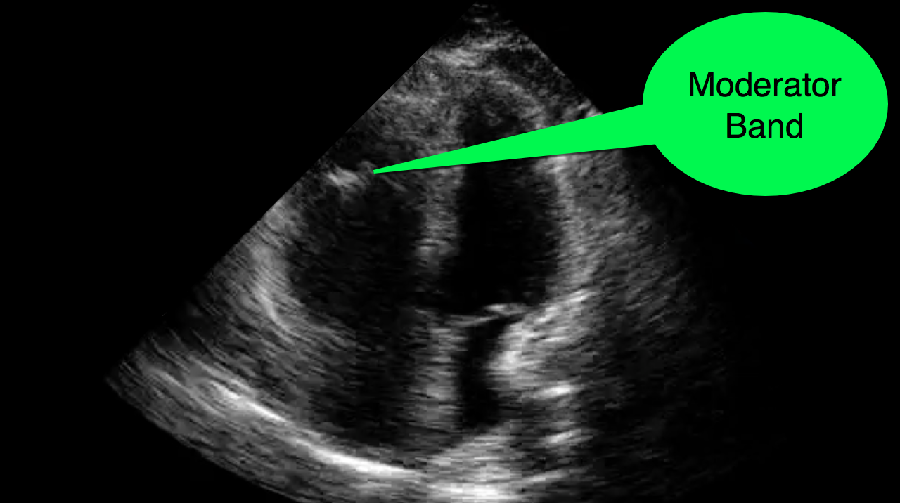
Then you can move on and be able to closer examine and evaluate the chambers and septa, moderator band, foramen ovale, a portion of the aorta, as well as the valves for any defects.
Radiopaedia does a great job on explaining the 4CH view and the possible defects you can see :
https://radiopaedia.org/articles/four-chamber-cardiac-view-fetal?lang=us
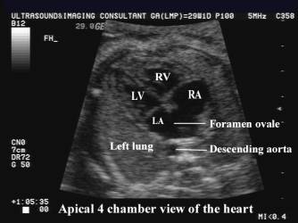
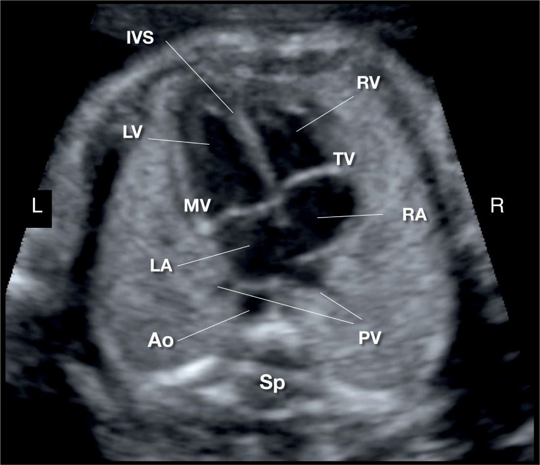
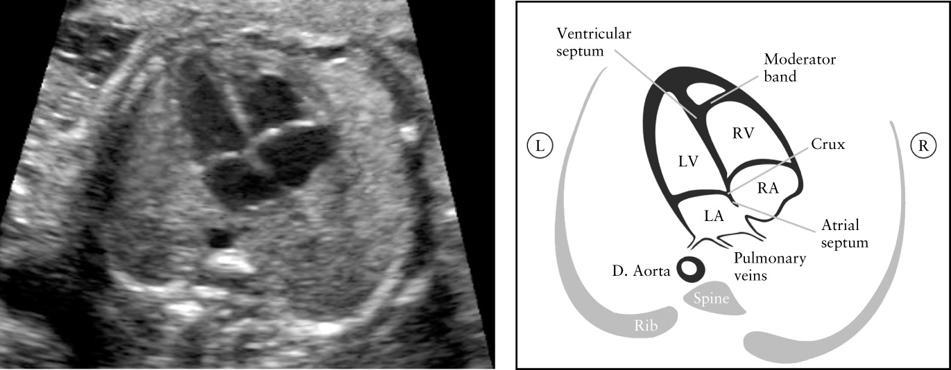
This is a great diagram of the 4CH view with corresponding sonographic image, and another sonographic image of apical 4CH:

Hi Karen,
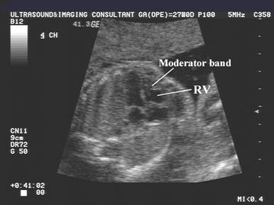
Great call to include the moderator band! It was interesting to learn that it will always be in the right ventricle, so that can help determine if the heart has developed left and right sides correctly. Apparently in the case of some congenital heart disease, the ventricles may be more ambiguous. Fun fact: It is also referred to as the septomarginal trabecula.
Hi Karen!
Great post! I love the images you added as well, it really helps me visualize all the structures I should be seeing. I feel like just since we talked about all of this yesterday, I've made more of a conscience effort to see all these structures while I'm scanning during clinicals!
To get the 4 chamber view of the heart, we should obtain this from the subcostal view or the apical view. In this view we should see the spine and one whole rib.
Things we should evaluate in this view include:
- Presence of four chambers
- Symmetry between the left and right chambers of the heart
- Shape
- Size of the heart (~1/3 of thorax)
- Position of heart and apex
- Intact septa (excluding the foramen ovale). Double check for any septal defects, especially at the crux of the heart.
- Foramen ovale. Can you see the blood flow through it, whether its from the flap swinging into the left atrium or with color doppler? Is it normal in size?
- Atrioventricular valves. Are they working properly? Are there signs of regurgitation? Is the tricuspid valve more apical than the mitral valve?
- Contractility of the atria and ventricles. Does the timing look correct and is it consistent? Are the ventricles contracting with the same force?
- Heart rate using M-mode
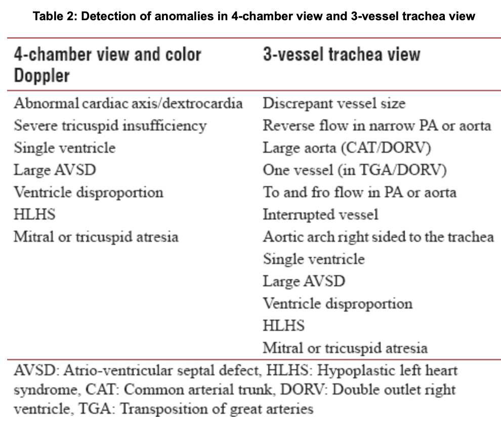
It is important that we perform a thorough exam of the heart to catch any abnormalities and set the fetus up for better management in the future. Below is a chart of the cardiac abnormalities we may detect in 4 chamber view and the 3 vessel view. The article linked below has great information about the fetal cardiac exam as well.
Great job Alexis, very thorough! What are some other things we can look at outside the heart at this view?
Thanks, Paris! Outside of the heart at this view, we can evaluate the lungs for homogeneity and echogenicity. We can also see the descending aorta posterior to the left atrium and anterior and a little left of the spine. Please let me know if I'm forgetting anything!
The importance of the four chamber view cannot be stressed enough. Within this single image there are many areas the sonographer should evaluate. It is at this level that we will view the heart's anatomical location and position. We know that the normal position of the heart is levocardia, with a 45 degree angle to the left of midline. We should also see the heart occupying about one-third of the thoracic cavity with anything greater than that indicating either a small thorax or cardiomegaly.
As we discussed as part of today's lecture, it is important to review the three S's of the heart. The three S's include the heart's size, shape, and symmetry of contralateral anatomy. The Atria and ventricles should be similar in size and we know that the heart should form almost a triangular shape in the chest cavity. In addition, we will also ensure the bilateral Atria and ventricles have perfect symmetry as we know there are some anomalies we're patients could present with a hypoplastic ventricle! Also, be sure to observe the moderator band found near the apex of the right ventricle, this is a great way to determine right from left.
We should watch a full cardiac cycle several times to make sure the heart is functioning properly. Observation should also include the opening and closing of the atrioventricular valves and the foramen ovale. We want to locate the interventricular septum to make sure that there is no communication between the right and left ventricles as this could indicate septal defects!
Here is a great site that breaks down fetal cardiac imaging with some awesome pictures: https://radiologykey.com/sonographic-evaluation-of-the-fetal-heart/
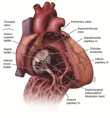
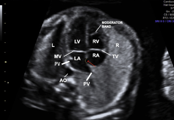
Here's a neat picture that gives a closer look at the moderator band!
Hi Chrishawna!
I really appreciate how you began your discussion post with the concepts from last week! Those concepts totally flew over my head and it's great that you went back to explain that the fetal heart should be laying more towards the left side of the thoracic cavity at a 45 degree angle. With all the classes and clinicals, it's easy to forgot how essential is its to know your basics to build a strong foundation for the rest of the quarter.... and the rest of our lives.
After completely bombing the quiz and then writing it out as an explanation for this discussion post, I will never forget that Three S's of the heart.
The images that you shared and your post was amazing! Thank you!! This really helped me out so much
Hi Chrishawna! I think you are spot on. I like that you mentioned we should watch a few full cardiac cycles to make sure all the mechanics of the heart are functioning properly. I'm wasn't really sure what normal looked like so I found this short video clip of a normal fetal heart beating in the 4 chamber view. I also found a second video showing a couple fetal arrhythmias just to see what some abnormalities would look like.
Normal fetal heart:
Common fetal arrhythmias:
Nice job bringing up the moderator band Chrishawna! That is definitely an easy way to orient yourself if you see it in the right ventricle! Good thinking with the hypoplastic ventricle if it is not symmetrical and similar in size as the opposite side. I did some searching on the survival rates of infants born with ventricular hypolasia; can you think of some things this type of defect may lead to in a newborn?
Hi Chrishawna!
I totally love how you incorporated the three S's. We definitely want to remember that when interrogating the heart and 4CH view. I also want to add that it's super important we see a solid interventricular septum and when we're scanning, get accurate pictures that aren't creating deviations or dropouts in the septum, because this could be a major problem if there truly is a deviation.
Hey Chrishawna!
I love that you acknowledge the importance of the moderator band, however, its not the best way to determine right from left. You haven't learned this yet, but there are pathologies that can cause thickening on the myocardium and it can almost mimic a moderator band. There are also pathologies that can make the walls hypoplastic. If there is anything I can instill in you guys it's that we shouldn't be using landmarks inside baby to determine right from left. It should be determined ONLY using fetal lie. Then once everything is definitely where it should be, then landmarks such as the stomach and the heart (left side) can be used.
Thanks for sharing your thoughts!
Hi Paris,
I am so glad you mentioned this! I think sometimes I get too hung up on landmarks to orient myself and need to pay more attention to fetal lie when doing OB or where I am in the body with abdomen. I think of landmarks as my safety net and hope they will point me in the right direction, but we must never assume normal no matter how tempting it may be to do so! :)
The 4 chamber view is one of my favorite fetal heart views. I love how you can see so much in one image!
In this view you can first determine the position of the heart, the axis, the shape and size, and also the symmetry of the atria and ventricles.
Next you can evaluate the right side of the heart and should be able to see the:
In the left side of the heart you should be able to see the:
You can also see the septums, lungs, at least one whole rib, and a cross section of the descending aorta and fetal spine in this view.
Hi Brittany,
Great job listing out everything we see in such an organized way! Your post is very clear and to the point, love it! We may all put your post in our notes.
That is a really good image you found, as well. I found it a little difficult to find one that has so many things labelled.
Thank you!
Karen
When evaluating the heart, the first thing we want to do is confirm cardiac activity, after this, assessing situs is the next thing we would want to confirm right off the bat. Then looking at shape, size, and symmetry is very important.
Then we can move on to the anatomy of the heart in this 4 chamber view. This includes:
Evaluating the right and left ventricle and the right and left atrium. The foramen ovale, mitral valve, tricuspid valve, moderator band, interventricular septum, interatrial septum, and muscular and membranous septa
The pulmonary veins and the descending aorta may also be seen
nice anatomy reminder!
Thanks for adding these pictures to go along with the DB. I was thinking about the mitral and tricuspid valve and was wondering if you would be able to see the three and two valves. I believe DR. Wilson said that the membranous septum is more common to have the defect than the muscular septum is? 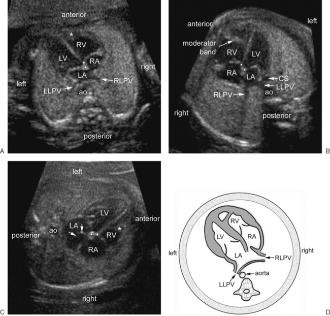
Great job mentioning the foramen ovale! Michelle was showing me a case at cinicals once and they could tell there was something off about the heart. There was nothing huge, but it wasn't checking off all her boxes. She told me to look at it. One of the first things I noticed was that the foramen ovale was opening towards the right atrium rather than its usual left! It was something so small, but it definitely makes a difference! It makes you wonder how that blood is even circulating!
So many things to think about...
Oh my gosh Paris, thats amazing that you caught it! I love when you share your experiences with us, they're always so interesting. Yeah that's definitely something to wonder about, the human body is so resilient even when something is off like that! Remembering all these small details is so important not only to remember for class, but we seriously need to use our knowledge from this class in our everyday scanning.
Hi Hailey,
Great job with the visuals, it really lists out everything that would be in the 4CH view! At this point, when we scan I'm sure we're just looking at a few key things, haha. But I love that as this class goes on and we see more diagrams like the one you shared, we are more and more likely to be able to list off the different anatomical structures that we see.
I'd love to see a fetal heart now and be able to list off some of the structures that we've learned just this past week.
Have you been able to scan any OB at your site?
Thanks!
Karen
Hi Karen,
thanks! These diagrams really help with repetition, seeing all the images the girls post is so helpful. we don’t get a ton of OB, usually first tri coming through the ED or A biophysical profile here and there. what I’ve been doing is while observing the biophysical profiles, I try and name the structures I see when they sweep around. I‘m not able to scan during those exams because of the time limit they have. hopefully I’ll get some second and third trimester practice in soon!
I know you mentioned that you don’t get a lot of OB either, do you think you our learning would click more if we were able to learn it, and then go out and scan it?
In my opinion the 4CH view is the easiest to get and most helpful in determining if there are any septal defects. You should be able to see the following.
This website had a great breakdown of what you'd what to see as well as some images and cases with this view!
https://radiopaedia.org/articles/four-chamber-cardiac-view-fetal?lang=us
Hi Lania!
Love your list, very organized and easy to read. you mention that we see pulmonary veins. Do you know which ones we most often see? I was getting confused because Dr. Wilson said inferior but in the recorded lecture she said superior.
Hey Lania and Hailey! We discussed this in our DB but I believe you see inferior and superior coming out of left atrium. Can someone tell me where the pulmonary arteries come out of. 
Great job mentioning pulmonary veins! We take those guys for granted because they are not always readily seen! But all you have to do is wiggle the probe around and they should come into view.
Awesome!
So sweet and concise! Thank you for sharing the break down and your resources, Lania!
Definitely adding this information to my notes. You think you understand something and more information comes at you! We should get together and break out a list for every other view lol. I'm finding this discussion post so helpful!
As a sonographer, it's important to sweep through and evaluate the heart in multiple scanning planes. We will ALWAYS begin the fetal complete survey with the evaluation of the heart to ensure that there is life. Assuming that we are scanning a 20 weeks old fetus, it is also important to investigate the 3 S's: size, shape, and symmetry! So as we continue our scan, we will get to the four chamber view of the heart. You will know that you are at an optimal level of view of the four chamber plane as you are also in view of the apex of the heart, the superior pulmonary veins and one whole rib. We would need to evaluate: the right ventricle that is behind the sternum and anterior towards the chest wall; the normal motion of the foramen ovale flap by the oxygenated blood flow from the IVC; that the right and left atrium forms the base of the heart as the right and left ventricle forms the apex of the heart. These views should help determine the position of the heart within the fetal chest. 
Howdy Monica,
That's a great way to remember the apex of the heart!
I think it's easy to overlook but the aorta is a helpful marker when looking for that 4 chamber view. As simple as it sounds, seeing the aorta can help orient left/right and anterior/posterior.
Sarah
Great response, Monica! Can you think of what we look for in the 4 chamber heart view if we come across a dextrocardia or mesocardia fetal heart to identify the correct atria/ventricles?
Dextrocardia, yes. because it'll just be angled the wrong way. Mesocardia.... no? Just assuming that its squished.
TY MS. LAUREN B
Great job Monica!
Lauren, I think Michelle mentioned we can look for the moderator band in the right ventricle to help us determine which ventricle is which? Correct me if I'm wrong though please!
https://umem.org/educational_pearls/2751/
Hi Zuly!
I love this cine clip you posted of the heart showing the moderator band within the right ventricle. I can’t remember if Paris and Dr. Wilson said we could rely on visualizing this landmark to confirm it’s the right ventricle or if sometimes it’s not accurate to go by. Would love any feedback on this from you Paris. Of course fetal lie is our goto for determining proper positioning but if we can also utilize on the moderator band to indicate position that would be great as well. Thank you!
One thing I always do when I am taking my split screen situs picture (heart in one image and stomach on the other), I always annotate "apex" on the heart image. This lets the radiologist (and myself) know that I'm certain that the heart is pointing where it should be and even at the appropriate angle.
The four chamber heart view is such an important view to obtain and should be the first heart image taken.
Along with the landmarks I'm going to list below, what should also be visualized in this view is 1 full rib, apex of the heart, interatrial and interventricular septum's, descending aorta, the 3 S's (shape, size (1/3 of thorax), symmetry), and finally the proper position of the heart being 45 degrees to the left of the chest.
Right side of the heart:
1.right ventricle
2.right atrium
3.tricuspid valve
4.moderator band
Left side of the heart:
1.left ventricle
2.left atrium
3.mitral valve
4.inferior pulmonary veins
5.foramen ovale

Hey Molly,
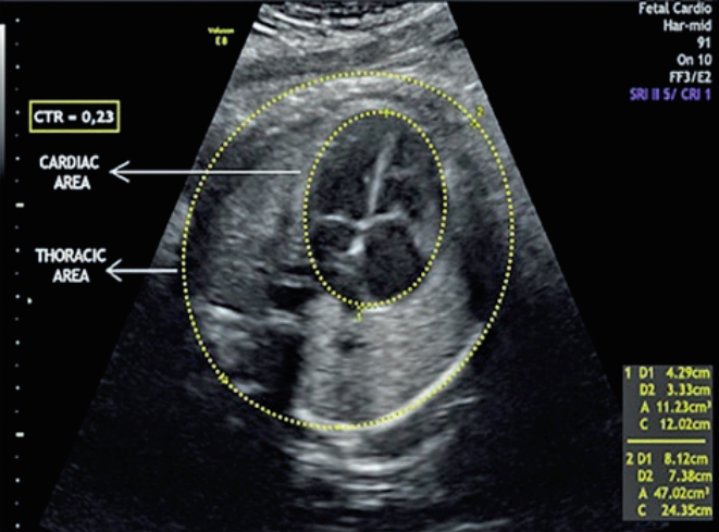
Great job our your response! One thing you mentioned that I found to be super important is the anatomical landmarks we should see when capturing this image! I noticed all of fetal biometry has specific areas where measurements should be taken so I assume this is also the level we could take the thorax circumference or cardiac-thoracic ratio as well!
Hey Molly! Your DB included everything I thought you should be able to see in the heart. Do you remember the degree variation the heart should be within when it is at 45 degrees?
SHAPE SIZE SYMMETRY!! I will always remember it now that I got it wrong on the quiz haha
Hey Molly girl,
I love how you divided your response by left and right side. I am so confused though about the differences between the interventricular septum and septum primum and septum secundum. Do you have an understanding on this?
Hi Nailah!
I love your post! It's great how you labeled everything so clearly. The images are super helpful!
I am also having a hard time understanding these septa's. I found this information online and it is seeming to make things more clear. I hope it helps you too!
The septum secundum: is a muscular flap that is important in heart development. It is semilunar in shape, and grows downward from the upper wall of the atrium immediately to the right of the septum primum and ostium secundum. It is important in the closure of the foramen ovale after birth.
Septum primum: is the division between the right and left atrium.

The interventricular septum has two parts: muscular and membranous
1. Muscular part
- Appears as a muscular ridge corresponding to the interventricular sulcus.
- It has two limbs and a free margin
- Posterior limb advances towards the posterior endocardial cushion
. - Anterior limb approaches bulbs cordis.
- Free margin of the septum guards the interventricular foramen.
2. Membranous part
- This part of the interventricular septum has multiple origins.
a) Posterior endocardial cushion b) Right bulbar ridge c) Left bulbar ridge

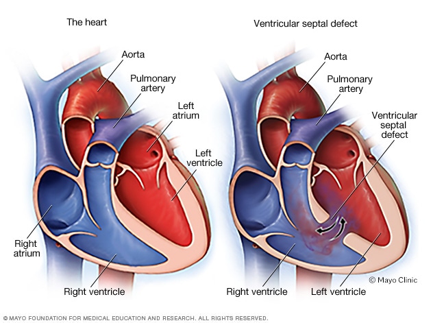
Omg thank you so much molly girl! YOU ARE AWESOME !!!!! This so organized and informative, definitely screenshotting this! I really appreciate you providing this addition information, mean a lot! I am thinking the septum primum and secundum must be the precursor for the interatrial septum like in first picture right?
Hey Nailah!
I’m happy you liked the visuals!!
I found this information online regarding the interatrial septum forming:
Soon after birth, the elevation in left atrial pressure forces the septum primum and septum secundum together, collapsing the space between the septa and therefore closing the opening between the left and the right atria. At this point, normally the septa will fuse and become the interatrial septum.
This definitely helped me. Thank you for your question!
When scanning the 4 chambers of the heart in ultrasound, I know you are looking for size, symmetry, and shape. You should also check for the situs of the heart with situs solitus being the normal (heart on left and pointing towards to left). If any of these look out of the ordinary then the measurement to ensure that the circumference of the heart is within 1/3 of the circumference of the chest cavity. You are supposed to evaluate the ventricles and atrium and check that they are equal but in the second and the third trimester the right side of the heart becomes slightly larger than the left.
The septum’s and valves should also be evaluated. The foramen ovale should be evaluated in between the left and right atrium and should be flowing into the left side. I am not sure if you put color or do anything else to it.
The tricuspid and bicuspid valves should be evaluated for functionality and proper contraction.
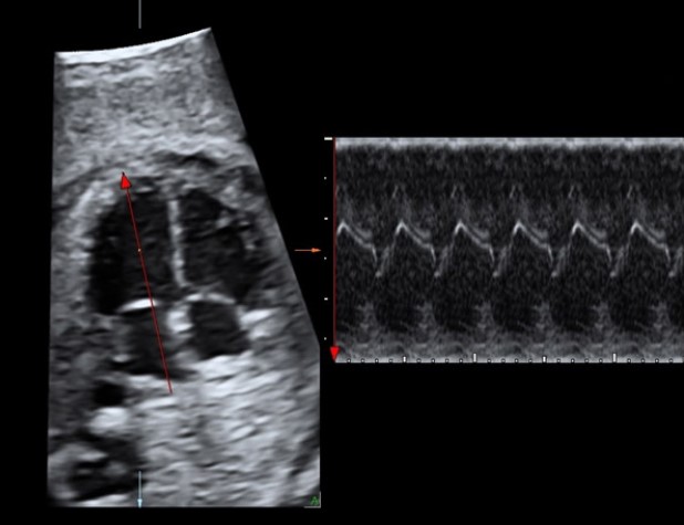
Heart rate should be taken using Mmode and should be around 120-160 bpm.
Ensuring that there is also nothing in the lung space is also important.
Hi Candee!
I noticed how you mentioned assessing the foramen ovale and I think this is definitely an important part of a fetal exam. We want to assess that firstly there is an opening between the ventricles, and that blood is able to flow freely between the two ventricles. If flow is impaired this can alter the fetal circulation system. The best way to do this would presumably be with color doppler and maybe M-mode too?
I also saw a few studies that were looking into measuring the foramen ovale opening and relating this to IUGR! Definitely seems like we may eventually do more in terms of assessing this structure.
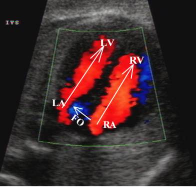
Here's an image of what the foramen ovale looks like with color Doppler on! You can see the flow entering the left atrium.
Hey Candee girllll,
Hope your day at clinic was good, really good response. I just wanted to add that we also see the pulmonary veins. I'm not sure which ones we see most often because Dr. Wilson mentioned both. Do you know? Inferior or Superior. We also see the interventricular septum! so many small parts that are too easy to forget about.
Hi Hailey!
Thank you for bringing the pulmonary veins up!! I had the same question as you about if it's typically the inferior or superior veins seen.
From Dr. Wilson's lecture I remember her explaining that in order to determine if you're in the right plane for the 4 chamber view you should see the apex of the heart, the superior pulmonary veins, and a rib. Page 515 of our textbook also states that the superior pulmonary veins should be seen in the 4 chamber view.
However, later in our lecture I believe it was stated that the inferior pulmonary veins are typically the ones seen.... Maybe it varies?
Hey Brit! I was looking into the pulmonary veins also. From all the image I've seen, the superior and inferior pulmonary veins empty into the left atrium pretty close to each other. I wonder if they're so close that maybe it's a tiny micro-movement to see superior vs. inferior. And if so, maybe the change in angle is so small that it doesn't affect the other components of our 4 chamber view that much. I'm hoping to check it out on the phantom in lab on Friday and maybe that'll help. I also watched this video that images and dopplers the pulmonary veins. The narrator didn't specify which PVs he was imaging. Perhaps seeing one pair in the 4 chamber view is enough?
https://www.youtube.com/watch?v=2nIocmxf1B0
Hi Alexis!
Thank you for the clarification and video! I agree with you that they're so close it may be hard to tell exactly which pair of veins we are seeing and it may be enough to just see one pair.
Hopefully you can get some OB exposure in clinicals soon and see if your CI has any information about it!
Hi Hailey, my day sucked thank you for asking. Hope your day was good friend. I didn't think about the pulmonary veins and arteries. However, we should see the superior and inferior pulmonary veins out of the left atrium and there are two arteries coming out of the right ventricle. I wonder if you see either of these during LVOT or RVOT please let me know.
For the interventricular septum we are just ensuring that we see the seperation of the left and right ventricles right?
Hi Candee,
Great insight! I'm still trying to wrap my brain around levocardia vs levoposition when establishing situs.
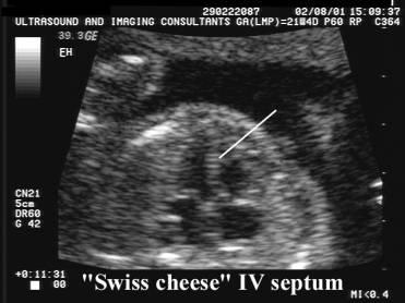
The septa and valves should absolutely be analyzed in the 4-chamber view. I was looking for more information about potential septal defects and came across what is referred to as a Swiss Cheese septum. This describes "uncountable" ventricular septal defects. Thankfully it sounds like it is quite rare. I'm sure we'll dive into all the different anomalies and pathologies next quarter, but it is interesting to start investigating now as we learn the normal anatomy.
Positions of the heart are very difficult to remember especially since there are different positions of the baby and it is hard to find the heart at all.
Levocardia heart of left apex on the left
Levoposition heart on the left apex on the right
Dextrocardia heart on the right apex on the right
Dextroposition heart on the right apex on the left
The way I can remember stuff is by grouping things together so I remember if it ends with "cardia" it will be both on the same side.. heart on left apex on left.....heart on right apex on right. Hope this helps.
Also, interesting defect to keep an eye out for. It is so small it's difficult to see.
Uh oh... I think I may have misunderstood these terms now that I see you explain them this way.
I thought terms like levoposition and dextroposition referred to the literal location of the entire heart. (levoposition meaning the heart is in the left side of the chest & dextroposition meaning the heart is on the right side of the chest)
And I thought the terms levocardia, dextrocardia, and mesocardia referred to where the apex was pointing? (left, right, or middle)
I assumed that terms like levocardia and dextroposition would just be combined to explain a heart in the right side of the chest but with the apex pointing to the left but this definitely may not be the case!
Do you have any references for this info so I can brush up on it more? Thanks for sharing this!!
Hi Candee, great job! Everyone has put such thoughtful responses to your post already, but do you remember why the right side of the heart grows larger than the left in later in the pregnancy? In addition, do you think if we don't see that shift in symmetry or if we see it too early, that could point to some issue in the fetal heart?
Great job noting the direction of the foramen ovale! I've had a situation where it was backward. I explained in an earlier post ^^
You don't need to put color on the heart unless you are further investigating something pathological or trying to prove a fetal demise.
The 4-chamber view is one of the more "iconic" images of a 20-week OB scan. This is where you evaluate the heart shape, size, symmetry, and situs. This is also where you check for fetal heart activity before proceeding with the exam.
I found this page on radiopaedia super helpful in understanding all the different things to look for.
Without going too far into abnormalities, this view is best for detecting septal defects, persistent truncus arteriosus, some hypoplasias, and the presence of any echogenic intracardiac foci (EIF). I observed an EIF on an OB scan last week. According to the protocols, I believe this is a "soft marker", and because the fetus also had a 2-vessel cord, it flagged it for perinatology.
This is what an EIF looks like:

Radiopaedia also notes that the 4-chamber view is used to establish heart location and fetal heart rate, as well as the atrioventricular (AV) valves -- tricuspid and mitral.
We typically don't even mention an EIF unless there are other soft markers as well. They are super common and are actually typically just the chordae tendineae reflecting the sound beam!
We will definitely talk about all those pathologies next quarter. What are some normal things we can be evaluating in a 4ch view (in the entire thorax)?
In the 4ch heart view we can also evaluate the lungs, as they should typically account for the other 2/3 of the thoracic space. The aorta should be visualized. I have also observed that the typical 20 week scan includes long/trans views of the spine at cervical, thoracic, lumbar, and sacral levels, so it would also be a good time to evaluate the 3 ossification points in the thoracic spine.
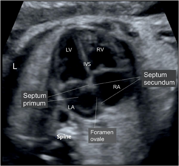
There are quite few aspects in the 4-chamber heart view that can be assess the development of the fetal heart. These areas include:
This diagram is really nice:
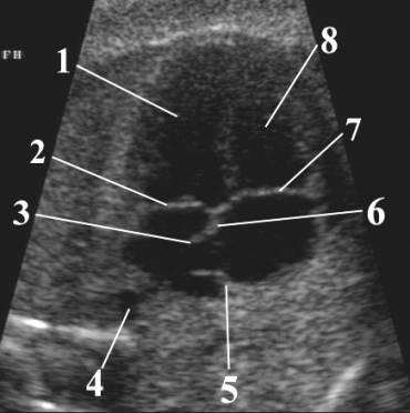
1 – Left ventricle
2 – Mitral valve (closed during ventricular
systole)
3 – Flap of foramen ovale
4 - Descending aorta
5 – Septum secundum
6 – Septum primum
7 – Tricuspid valve (closed during ventricular
systole)
8 – Right ventricle
aahhh and the pulmonary veins I totally forgot!! There was some confusion though if the veins in this view were the superior or inferior ones.
Howdy Class,
There are a TON of things we can learn from and see in a 4 chamber view.
All four chambers, their size, position, contractility
The heart's echotexture/echogenicity, location in the chest, size compared to the rest of the chest, overall symmetry
Moderator band band, septum, existence of pulmonary vessels, AO.
Bi/tricuspid valves
Heart muscle thickness
We love Radiopedia.
https://radiopaedia.org/articles/four-chamber-cardiac-view-fetal?lang=us
Sarah
Yesssss the muscle thickness! This is one of those things that you get so used to seeing normal that when the muscle thickness is off, you can tell! Awesome thinking!
Great job on your post Sarah!
I hadn't thought about the heart muscle thickness, thanks for mentioning it! I found a few images that might be good examples of how this can appear on this webpage.
https://obgynkey.com/fetal-cardiomyopathies-and-fetal-heart-tumors/
When scanning the 4- chamber view of the fetal heart there are many things that need to be visualized.
1. Situs
2. Axis
3. Size
4. Heart Rate(M-mode)
5. Atrium
6. Ventricles
7. Atrioventricular Values
8. Crux of the heart for possible defects
I like this image because it has a lot of labels!!!
Hey Allison,
Nice response. I really appreciate the organization. I like how mentioned the crux of the heart too! So important! I got confused on the foramen ovale. The lecture said the flap should be seen within the left atrium, but other sources are mentioning it is between the both atria and this eustachian valve plays a role? The book super confused me on fetal circulation. I thought I had a mild understanding, but maybe not.
nailah
Hey Naliah,
I think the eustachian valve plays a role but is more involved with the IVC and the right atrium. From what I understand the eustachian value sort of pushes the blood from the IVC to the foramen ovale and the foramen ovale flap should open and be seen opening in the left atrium. Here is an image of where the eustachian valve is located. In this image the foramen ovale is not fully formed yet.

Thank you so much Allison this is super helpful for sure !! I truly appreciate this information. Makes soo much more sense now. So then the forman ovale flap is not necessarily in the right atrium, it is the middle of the right and left atrium and opens into the left atrium when this valve is pushing oxygenated blood through?
Hey Nailah! From my understanding, you got it. The foramen ovale is an opening in the septum between the right and left atria and the flap swings from right to left when oxygenated blood comes in from mom. If you skip to 16:37 of the fetal echo lecture linked below, it talks about the typical movement of the foramen ovale and has a video of what it should look like.
https://www.youtube.com/watch?v=2qfGzNh2dR4&t=2s
Love how this is organized Allison, great job!
I just want to clarify two things:
1- you can't clear situs from a 4ch view. This as a radiologist. If they are just given an image of the 4ch heart, that doesn't tell them how the fetus is lying and if it's situs solitus (normal situs). We can only clear situs based on fetal lie. I think I know what you were getting at, but I just wanted to make sure we are on the same page.
2- When it comes to the crux of the heart, you're right, that's SUPER important! Like Michelle said in class, sometimes based on how the baby is lying, you can get drop out and that's typically where it occurs. We as sonographers have to make sure we aren't creating pathologies with artifact. When taking 4ch images, be sure there is no drop out right in the center!
Great job!
Hey Paris,
Thank you. Yes, I get what your saying. I meant more so just having the knowledge of where the heart is so we know what we are looking (which ventricle, etc) at if the heart is in a different spot on the body. Haha just how my brain has to process this organ!! But yes, totally wouldn't show anything in the typical 4-chamber view.
Hey Allison,
I love the way you organized your response. You mentioned something that is so important, but that I feel would be easy for a student to overlook, and that's the opening of the foramen ovale. It's hard to imagine an organ the size of a pocket change could have that many key structures within it. It got me wondering what would happen if the FO didn't form properly or never formed at all? I guess blood would have no choice to go through the right ventricle and through the pulmonary artery!
Allison Wine always on the beat!!
Wow your discussion post is gold. I won't be surprised if I pick up the next edition of a Penny book, your name is going to be on it.
I appreciate how you created this into sections. Taking little bits of information from this huuuuuuuuge concepts is helpful.
Looking at your's makes me realize how much information I missed on my post. Especially the the area of most frequent septal defects, the crux of the heart. But also M-Mode! Thats super important to take note of during a fetal scan.
It's interesting to know that almost 90% of the time, if the baby has a higher heart rate...its a girl & if the baby has a low heart rate... its a boy!
Hi Allison!
Great response and I love the image you posted!
All super organized and well explained! I also wanted to add that the apical view of the heart is what most radiologist want to see for our 4ch view of the heart when doing the exam!
Great response Allison! Like Paris and Michelle said, at the crux and interventricular septums of the heart there can be drop out due to the baby's positioning and ossification points of the spine. What are some ways you can overcome this artifact and how do you tell the difference of an artifact versus an actual abnormality?
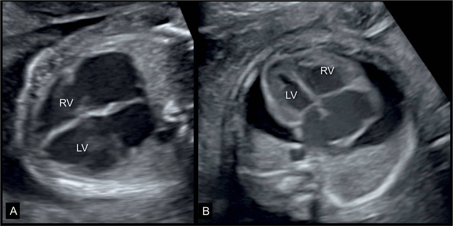
In the 4 chamber view many structures and factors may be evaluated. We want to make sure we see a complete rib in this view in order to ensure we are in the correct plane.
These may include the cardiac position and axis, the shape, size, and symmetry of the cardiac structures, the apex, and size of heart in relation to the thorax.
Left atrium, left ventricle, right atrium, right ventricle.
Foramen ovale, tricuspid valve, moderator band, mitral valve,.
Interventricular septum, inferior pulmonary veins, part of the descending aorta, crux of the heart, fetal heart rate using M-mode, and we may also evaluate the lungs in this view.
I liked this image since it demonstrates the plane/level at which we are cutting the heart in when we obtain the 4 chamber view!
Hi Zuly!
Great post and image! I just wanted to say I appreciate how you brought it's important to see one solid rib. I can't emphasize how many times my CI has told me that while scanning lol. It's very important for an accurate image, shape, and size of the heart.
Hi Zuly!
Like Lania said, great job mentioning the addition of seeing a complete rib to make sure we are getting the correct view/ angle!
I also liked your diagram and how it shows the transducers' view "slicing" through the heart to get that optimum picture. Sometimes we talk about things but don't or can't really visualize it, so your post really helps.
Thanks!
Karen
Hi Zuly,
I love this image! I was wondering if you have been able to visualize the pulmonary veins at your clinical site. It seems like it's hit or miss seeing those important structures. I included a color Doppler picture that i think highlights them nicely, I hope to get good enough at OB to get images like this one day!
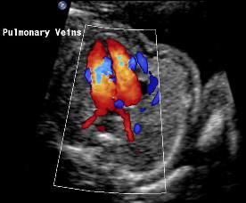
Thank you so much Crishawna!
I love this image, it demonstrates the pulmonary veins beautifully. I asked one of my techs today why we don't seem to get anatomy scans, just AFI, growth, 1st trimester and what not, and apparently they send those patients to a a neighboring location they call the foundation. I'm not completely sure how this works or why we don't scan them instead, but I suspect this is why I have yet to witness or scan 4 chamber view in clinic! I'm going to work with my techs to hopefully still be able to practice scanning these important views though!
When scanning a 4 chamber heart, we have to pay close attention to the size of the 2 atriums and 2 ventricles and see that they are roughly the same size (sometimes the right size maybe slightly larger due to increased workload). We need to make sure that the septums are separating the different chambers and that the valves are working efficiently. We need to also check the heart rate using M-mode to make sure it is between 120 to 160 beats per minute. We need to check if the heart is positioned correcting on the left side of the fetus and at a 45 degree angle and only occupies roughly 1/3 of the fetal thorax. If we have any doubts that the fetal heart may be too small or too big then we must measure the circumference of both the fetal heart and complete thorax and check the ratio between the two. The moderator band should be visualized, which helps us determine where the right ventricle is. You can use color doppler to see color flow crossing from one side to the next as well.
I think this website does a good job in breaking everything down. https://www.obimages.net/free-chapters/normal-fetal-heart-ultrasound/

Hi Raman!
Great job bringing up M-mode and the fetal heart rate! I feel like I need to look into M-mode a bit more as I haven't learned as much about it as we have Doppler.
Have you seen your CI use M-mode on the fetal heart much yet? If so, what view do they typically take the heart rate in? Do they only use it for heart rate or do they also use it for other purposes like chamber size?
I also found this cool graphic that breaks down what's being seen on an M-mode strip in this view.
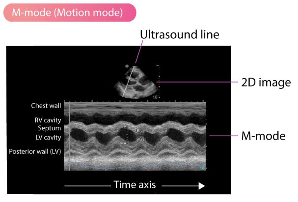
Hey Brittany,
Great image! I know we missed the typo on this quiz, but M-mode should be taken at an atrioventricular septum (between one atria and one ventricle).
This image is between two ventricles which doesn't help us determine how the atrium are pumping. This one isn't labeled, but I hope it helps!
Hi Brittany,
I like the graphic that you included in the picture. Yes my CIs have measured the heart rate using M mode just in the regular 4 chamber. I have also done it myself actually and they told me get the heart in 4 chambers and just put the marker anywhere? I honestly was so excited that I did not ask many questions but they could have just told me to place it anywhere since they know it's all new to me currently.
How exciting you got to do an M-mode on a fetal heart! That's awesome. Yeah, I'm also curious how specific the location needs to be for the marker. Like Paris said, I think as long as the line goes through an atrium and a ventricle you can get a good measure of the rhythm? Hopefully we learn more about this as I know M-mode has a few different functions!
Hey Raman,
No need to use color doppler! We really only need to use it if we think something pathological is happening or if we are trying to confirm a fetal demise.
I actually have a question about color in OB scans! I forgot what part we were imaging, but on a healthy OB patient my CI told me not to use the regular color doppler, but to use the PDI button, which is power right? Isn't that the one that we were told not to use since it has higher powers and therefore more heat sent in to the body? I didn't want to question what my CI did especially since I wasn't sure if that was the same thing, but the clarification would be nice if you have any :)
Thanks!
Raman,
The right side should have a little thicker myocardium for sure. Remember that we can learn all sorts of things from the 4 chamber heart view. Remember each outflow tract, the valves, AO, and the moderator band too. Also the septum from this view can give us some helpful information.
Sarah
Awesome responses so far everyone! It looks like we have some really great lists of what we need to look for, so now maybe we can use this to create some ideas for our circulation homework assignment. What are some interesting parts of specific cardiac anatomy that may trigger an idea for our classes stories? For example, I picture the aortic arch (even though not in the 4 chamber view) as the lead up to a big rollercoaster drop, and then where we travel straight down after the drop! Are there other parts with special appearances or pathways that may spark a story?
Hi ladies,
In case we didn't have enough images, here's another great one:
And here is a link for the ISUOG guidelines for 4CH view!
https://obgyn.onlinelibrary.wiley.com/doi/10.1002/uog.12403
Great work on the board this week everyone! I think we have a really good, cohesive list to study from now and we can hopefully reference this for our homework this week too. I think we need to remember that, as sonographers, we will not always be seeing perfect anatomy or scanning a healthy fetus, so we need to document as much as we can, as best as we can. If something is off in size, shape, or symmetry, or a landmark is not present, we are the first to document it and therefore aid in / lead to a treatment plan for the pregnancy and after birth.
Site pages
Current course
Participants
KPSAHS
Week 1 1/4-1/10 Role of Sonography in Obstetrics
Week Two 1/11-1/17 Fertilization and The Use of U...
Week Three 1/18-1/24 The Fetal Environment: Umb. C...
Week Four 1/25-1/31 2nd Trimester Evaluation &am...
Week Five 2/1- 2/7 Fetal Thorax
Week Six 2/8-2/14 Fetal Head and Face
Week Seven 2/15-2/21
Week Eight 2/22-2/28 Fetal Abdomen and Urogenital ...
Week Nine 3/1-3/7 Fetal Skeletal System
Week Ten 3/8-3/14
Week Eleven 3/15-3/21
Week 12 3/22-3/28
