Cervix measurement is definitely a tough one both transabdominally and on EV!
Today one of my techs and my CI had a discussion about this very topic, in regards to a scan I observed on a 3rd trimester OB patient. She had a cervical length order, and on EV it was immediately evident she had cervical funneling. No matter where she placed them, it was on the shorter end, but depending on where she put them, it would make a difference between lower limits and definite short cervix.
My CI said we should have applied pressure with a hand, on the pelvic region, while performing the EV scan, to see how it affected the cervix. I'm not 100% sure what information this would give us, but I thought it was interesting. In the end my CI said the cervix measurement, particularly the caliper placed on the external os, is somewhat subjective, but to be mindful not to extend it into the vaginal area.
https://radiologykey.com/uterus-and-cervix/
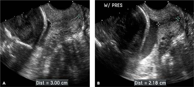
- The length and shape of the cervix may change during the course of the sonogram. This can occur spontaneously (Figure 17.1.4) or may be elicited by manual pressure on the uterine fundus (Figure 17.1.5). In either case, the likelihood of preterm delivery correlates with the shortest length of the cervix during the sonogram.
- Cervix dilating with fundal pressure .A: Sagittal transvaginal view of the cervix demonstrates a normal appearing cervix measuring 3.00 cm in length. B: With manual pressure on the uterine fundus (W/ PRES), the cervix shortened to 2.18 cm in length.
