Let's use this thread to bring up discussions on items we have needing more attention. Feel free to post up items that seem unclear or maybe you can add information to your peers on how you best understand some of our more complex items....
Week 11 Discussion Board
Hello beautiful ladies!!!
We did it guys! We are almost half way done with this program!!!!!
Thank you to Dr. Wilson and Paris for being such great teachers and helping us lay down the foundation for OB. We can hopefully impress the new teacher with our vast knowledge.
I was very intimidated at first about this class because of how complex it is and how the babies future is basically at the end of our fingertips (or probe I guess in this case). It is critically important for us to make sure we understand every little concept we learned in class. Let's use this board to help each other out like we always do and share some of our thoughts.
Lets go SonoSisters!!!!
Hi friends!
I think something that I struggled with in the beginning of this quarter was fetal presentation. However, I feel as though I've been able to come up with a game plan of how to asses the fetal position that has been making this task easier. My experience is mostly with the biometrics simulator in lab, as I haven't had much clinical experience yet, so please add anything your CIs may have taught you!
Steps for determining fetal presentation that have helped me:
Assess the mother's cervix - Is the baby's head there? Is their foot? Are they sideways across the cervix?
-
- if their head is at the cervix = baby is vertex / cephalic
- if their feet are at the cervix = baby is breech
- if baby is laying sideways = transverse position
Assess fetal spine in an axial view - transverse plane for breech/cephalic; longitudinal plane for baby in transverse position
-
- spine on top of screen = baby laying on tummy
- spine on bottom of screen = baby laying on back
- spine on left / right of screen = baby laying on their side
Combine info on where fetal head and spine are to get a full position of baby
-
- examples:
- fetal head at cervix, trans spine on top of screen = baby laying on tummy, head towards mom's feet
- fetal head at cervix, trans spine on bottom of screen = baby laying on back, head towards mom's feet
- fetal feet at cervix, trans spine on top of screen = baby laying on tummy, head towards mom's head
- fetal feet at cervix, trans spine on left side of screen = baby laying on its left side, head towards mom's head
- examples:
If the fetus is on their side I've also found it helpful to sweep through them coronally to help determine which way they are facing. If you are sweeping one direction and see an elongated spine you know you're at the back of the fetus and if you start sweeping the other way and see their face you'll know you're at their front. This has been particularly helpful for me with transversely positioned babies.
Looking at the organs can also be helpful to determine position but we must be careful with this and only use this as a tool if the fetus has normal situs. If it is normal then the heart and stomach should both be located on the left side of the body and the apex of the heart should point to the left side of the chest as well.
Let me know if you all have any other tips or tricks for this difficult topic! :)
Hi Brittany,
This posting is AMAZING!! Part of me wants to print this out and add to my clinical binder as a cheat sheet, is that weird lol? This has been one of my biggest struggles is getting fetal lie down! Not to mention the baby is forever moving and changing position which just adds to the confusion. One of the technologist at my site recommended drawing out the different fetal positions by using a transverse view and showing how the spine is in a different position depending on how baby is laying, I think I will try that this week!
Hi Brittany,
Awesome pictures in your post! I love how you broke it down starting from the cervix and then analyzing what you have on the screen. It is like putting a puzzle together. For me, I used Dr. Wilson's tip and used my teddy bears one as mom and the another tiny one as the baby and would practice like that. The CIs at my site also like you said do a quick sweep to check where the spine and head are and get a good idea of the baby's position.
Hi Raman,
Yes I find doing an initial sweep to be so helpful!
Today at clinicals we had a 20 week fetal survey which started with the fetus in a cephalic position and ended with them transverse! They were dancing around the whole exam haha. Definitely makes it a challenge and I have to keep reassessing how the baby is positioned.
Thank you so much for sharing this Brittany!
This + that afternoon where you and Allison explained in lab with the paper doll how to determine fetal presentation, really helped me. I agree with Chrishawna, this is a great thing to have in our binders for clinic, I actually already formatted it into a word document! (:
Such a great reminder!!!
I don't think I will ever forget doing this in lab together! I think the coronal sweep noting where you are pointing the transducer on moms belly as you see the fetus spine to fetus belly then following up with a transverse view looking for the location of the spine in correlation to the screen might be a fool proof way of figuring out fetal lye. By the way this is much easier to explain in lab while actually doing it.
Hi Brittany!
I just wanted to join everyone and say that this is a great post! Are you sure that you're struggling with this concept? You look like a pro!
I think, maybe its just being exposed to many OB scans, will bring you a lot more clarity! You're about to be killing it for your first hands on OB!
Your post has definitely helped me. I comprehend things in little bites, so I GREATLY appreciate this.
Thank you so much!!
I'm hoping I'll slowly get some more OB cases in clinicals. I did observe a 20 week fetal survey and today I got to scan an 8 week gestation! It's definitely a lot harder to determine the position when the fetus is moving around so much but I'm hoping to use these guidelines as a starting point!
Hi Brittany!
Great post my love!
I also have had an issue with fetal presentation. I love the tips and game plan you have to master it. I've noticed that imaging baby's fetal and then immediately followed by situs helps me get baby's position in my head easier. I also have to act as if I'm in the mom's belly sometimes lol to fully get the presentation.
I think most would agree that heart views pose a particular challenge, both conceptually and sonographically. I'm slowly getting a little better at understanding everything. I feel like with each exam I see, I gain a bit more. I also know that I need to oversaturate my brain with this concept until it becomes second nature.
Thankfully we live in the age of the internet!
I really liked these videos from Philips Healthcare that go over each of the views we obtain.
I'm also watching from Dr. Roy Filly. It's a longer video (I recommend watching at 1.25x speed), but I feel like hearing it explained a multitude of different ways will help make the information "click" in my brain.
Everyone learns material differently, so I would love to see what other resources you all have found to explain and conceptualize the fetal heart!
OMG LEAH! I love Dr. Filly's video and shared it in my post as well. I think it helps to keep watching it over and over and I just answer his questions before he tells the answer and pause the pictures that he has and try to see what structures I can see correctly.
Let's say you are scanning a baby and cannot get all the heart views you need, how do you go about this? Would you try to get all the heart views at once or move on and come back?
I would certainly try to get all the heart views at once. However if one was eluding me, I would still take what I could and move on. I've seen techs have the patient roll on their side, roll on their other side, lie on their stomach for 5 minutes (if physically possible), go empty their bladder, or walk around for a while, all to get baby to move.
Hi leah!
Wow thank you for all these helpful videos! I definitely am still getting the heart views down. I feel like I struggle with it because I don't get OB at my site. So sad. A patient was asking me things today and I told I wish I was getting more exposure to OB because it's what I'm learning right now. but there will come a time! Are you getting used to heart views at all in clinic?
Hello ladies!
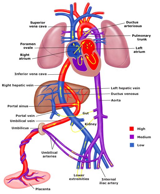
The topic that I'm mostly still struggling on is probably trying to solidify the cardiac pathways in my brain! I understand the concept of it but I'm still having a hard time memorizing the pathway. I've drawn it out and my brain for some odd reason still won't retain it lol it's literally rejecting it. So if any of you lovely ladies have any suggestions or tips or how to really get that down pat, I would greatly appreciate it! :)
This was a bit helpful for me!
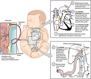
Hi gorgeous,
For me I think repetition is key and helps to make the material we learn second nature. I like to use the website slideshare; it has really nice presentations made by people that are simple and easy to follow. Here are two that I liked the most on fetal circulation:
https://www.slideshare.net/binuenchappanal/fetal-circulation-124643432?qid=f812c075-3760-465e-ae73-e3ba9c22ac64&v=&b=&from_search=10
https://www.slideshare.net/arvindyuvaraj/fetal-circulation-79357601?qid=f812c075-3760-465e-ae73-e3ba9c22ac64&v=&b=&from_search=6
I love NerdNinja too so here is short video of his that I watch occasionally to refresh this topic: https://www.youtube.com/watch?v=z1r6N168faw
As sonographers our job is to document images so knowing the outflow tracts and getting heart vessels makes sense, but why do you think it is important for us to know the blood circulation of a fetus?
Hi Love!
Thank you so much for these references! I'm definitely going to check them out ASAP! To answer your question, it's important we know the blood fetal circulation so we can make sure baby's heart is functioning properly. If we're not familiar with the flow, then we can easily skip past something worrisome during a scan and cause more damage for that baby by letting it go unnoticed!
Hi Lania. There is definitely a lot going on with the fetal circulation and it was confusing me at first too. What I found helpful was nailing down normal non-fetal circulation first since it follows a more straightforward path. I watched the video below a couple times to lock it in. Then I incorporated the additional fetal circulation pathways on top of that, like the foramen ovale, ductus venosus and such. I know everyone learns differently, but I hope it's helpful!
Hi Lania!
Not sure if anyone has shared this video yet:
It's from the Khan Academy and they always do a great job of breaking things down into a digestible format. This one really helped me.
Hey Lania,
I hear you gal, fetal circulation is not easy at all. I think these graphics are great and should further help solidify you understanding. If drawing is not helpful, I suggest getting a white board or blank sheet of paper and writing it of memory and correcting you mistakes with red and then repeat until you dream about fetal circulation ; ).
While there are certainly many concepts I have struggled with, I would say my top two would be determining fetal lie and how to obtain the heart views.
This week I plan on bringing a doll with me to clinicals. I understand the basics about cephalic vs breech vs transverse lie, but struggle when it comes to determining if baby is lying on their side, belly, or back! I find I understand more when I am the one scanning rather than observing, but it's still tricky and takes a lot of coaching myself through it.
When it comes to the heart I feel fairly comfortable with the anatomy and physiology, but I am struggling to get the LVOT & RVOT views consistently. I seem to have a lot of dumb luck with these views and can't always acquire them intentionally. One thing I have tried to do to help me was print out a few diagrams that show how to angle the transducer to get each view, so far it hasn't been super successful, but it's a work in progress!
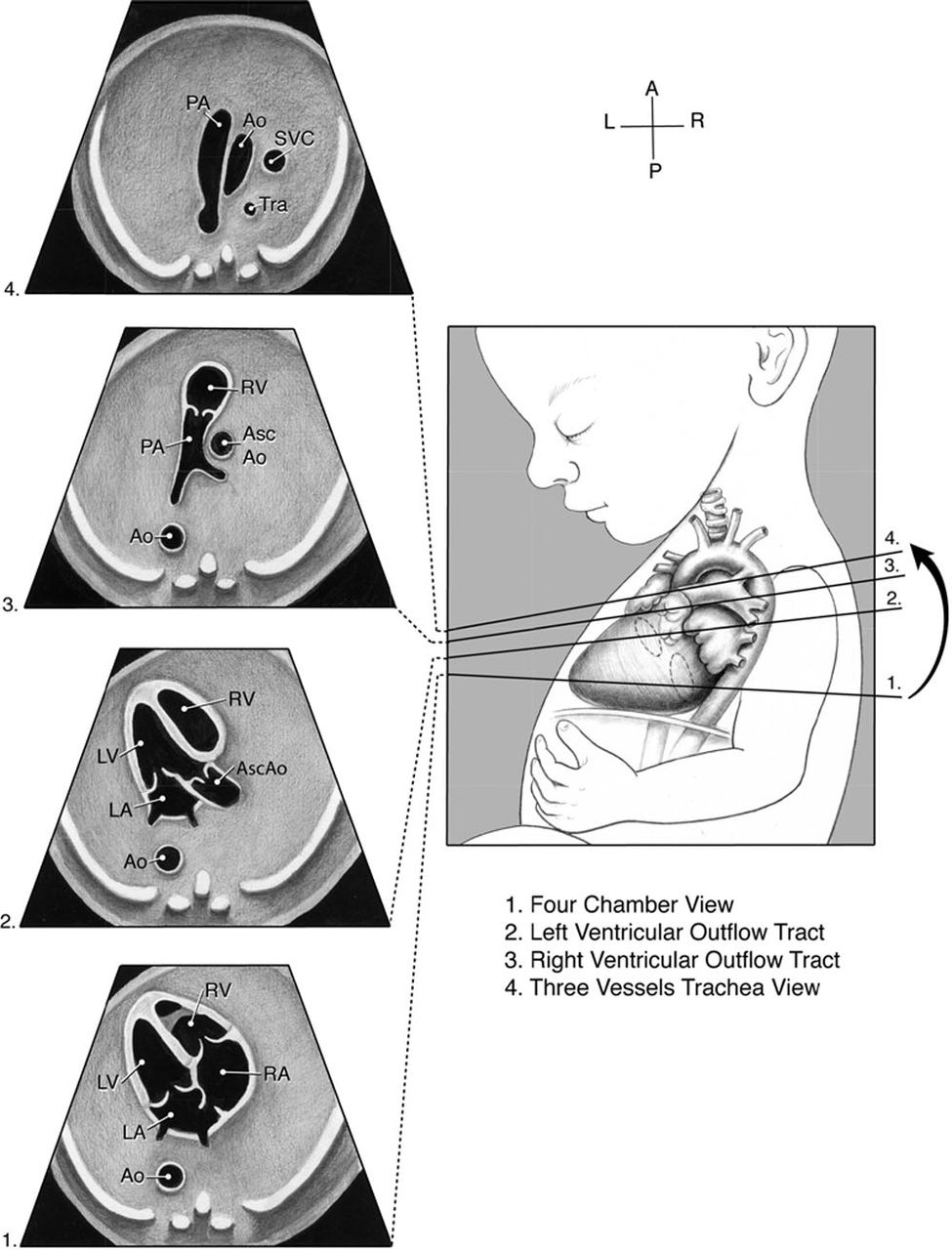
Hi Chrishawna,
I mentioned this earlier in another post but I used my teddy bears to help me with fetal postions :) It is a fun and cute way to learn these difficult concepts. I struggle with the outflow tracts as well and always try to see them as I watch my CIs scan OBs. I think the picture you used shows a bit how to angle our transducer up and down to get different views. Here is a website that shows the views we get and how one should angle their transducer to get those views: https://www.ultrasoundpaedia.com/normal-fetal-heart2/
When scanning OB cases, do you try to go system by system so get all the heart views all together or do you think its better to just get what you can and then move on?
Hi Raman,
Such a great idea to bring a teddy bear, I was looking all around the house for something and stumbled upon on of my dogs toys that's just the right size! Today was the first day I brought it to clinicals and it definitely helped me. I also found it helpful to draw the heart on it with a sharpie and the spine down the back!
When I have the chance to scan OB's I honestly try to go by system, my brain needs some sort of organization but it's easy to get off task if you stumble upon a view incidentally that may normally be tricky! What do you find works best for you?
Hi cutie,
Usually what I do is get a quick sweep of everything to make sure I know where the baby is lying and then I will go in and start from cervix to whatever is closest to the service. For example, today I scanned a baby who was in the cephalic position so I got all my head views first and then went to the abdomen and then heart. I know I lose focus too sometimes and I feel that since everything is new I hope to get all the pictures at once but that rarely happens. Today one of the techs and I were talking and she said as new students we tend to focus on just getting the picture but what is more important is understanding why exactly we are looking at that certain part. For instance, we are checking the heart for any abnormalities and making sure there aren't any issues with the septums or valves, etc. It seems like a lot but we will get there soon....hopefully end of this year :P
Hey Chrishawna,
I am with you girl! I struggle in the same areas as well. What kind of doll are bringing? I was thinking about doing the same thing because I am really struggling as you know from us practicing this past Friday. I just don’t want to bring anything to distracting for my tech. I honestly don’t get much practice at all getting heart views so I don’t have much advice to offer except that I know the moments are very subtle, to where the mother can breathe and it can change the view. Maybe these videos can help:
https://m.youtube.com/watch?v=rW2r55AS3Xk
https://m.youtube.com/watch?v=jDl_XpGUJEU
One day the heart will be a breeze for you gal.
Hi Nailah,
I attached a picture of my "doll" it's actually one of Bentleys toys he doesn't play with (you can see from the picture he has more than enough toys lol) that I am using as a makeshift baby. I only use it when I am behind the tech observing because you're totally right about it being a distraction! One tip a technologist gave me to obtain the RVOT is to find the fetal profile (mid sagittal view) and scan inferiorly to the heart and it should be in or near the RVOT view! Hopefully this will help!

I don't know, Bentley kind of looks like he wants the baby back now! Lol
Thank you for sharing your CIs way of finding the RVOT. I haven't been able to attempt many heart views at my site yet because she always has me start at the top of our protocol and I run out of scan time before getting there! I really hope next quarter I can switch up the order we start in so I can get some more experience with those; they're a difficult concept for me.
I'm sure someone has probably posted this in one of our other heart discussions, but it's such a good image to quick reference the planes we get heart views in!
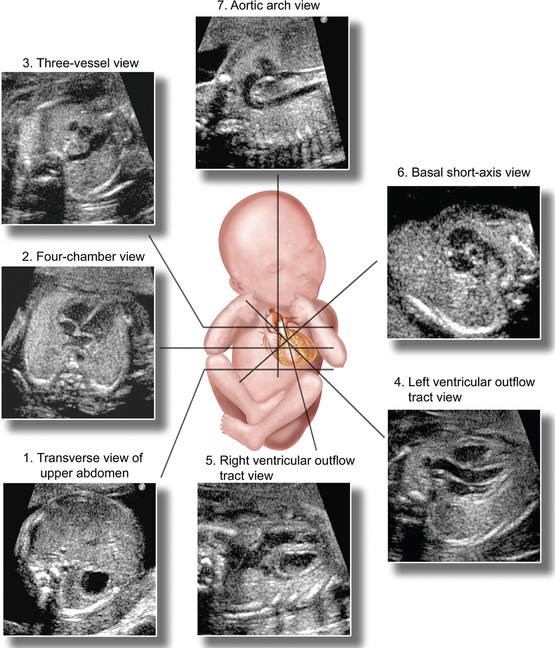
Hi Lauren,
He totally wanted his toy back now that I'm interested in it lol! That's awesome that your site is getting you used to their protocol, hopefully it will help you lock the images needed into memory so you never forget anything :)
Every week I try to concentrate on one new thing + 1 thing I struggle with (which is still basically everything). I feel ok about the cranial anatomy so I've been focusing more on heart and spine this week.
Thank you Chrishawna,
I am definitely on the look out for a doll. Thank you so much for those tips, when I do get a chance to scan fetal heart I will indeed try that. Here's a video I video I found with tips of getting a good four chamber view of the heart. He kind's of monotone but I find different methods helpful as I am still developing my own.
Hi guys!
Last discussion board post :(
I would say the topic that is still a challenge for me is embryogenisis and what structures develop from the midgut, hindgut, and foregut. I also am having a bit of trouble remembering when they develop even after watching the lecture a few more times.
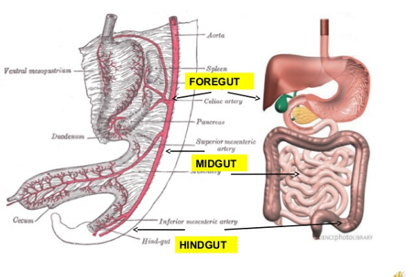
Hi Candee,
Ugh embryology is definitely something else lol. Here is a cute pnemonic that I found that you may find helpfu:
- foregut : """ BILL & PEGGY ON LSD "" (P=pancreas. E=esophagus. G=gallbladder. L=liver. S= stomach. D1,2 = duodenum 1 and 2nd parts. BILL = Biliary apparatus.
- midgut = JAILED CAT. ( J=jejunum. A=appendix. il=ileum. D=duodenum 2,3,4 parts. C=cecum. A=asc colon. T=trans colon prox 2/3.
- hindgut = DESCENDING RATS.
http://www.usmleforum.com/files/forum/2008/1/285338.php
Sometimes it is easier to learn a concept if we start from the end results rather than the beginning. So, what issues do you think can arise if the GI tract does not divide and form correctly?
The area I'm having some trouble with is fetal circulation! I wish I had time to stay after class today to go over it more like Dr. Wilson offered, but I had an appointment :( I found a few good videos though that have helped me review it since our circulation story assignment, and I'm getting there. I understand everything within the heart and from the UV to the heart, but I get mixed up when we start going to head, SMA, SMV, body. Here's one of the videos I liked that broke it down really well and showed a simple version before adding in each part and their names, so that we see the direction of flow first before adding in specifics!
Hey Ladies,
I have a a lot of trouble remembering dates. I think I am going to compile a list or a chart with all the important dates and measurements. If anyone has any way they remember these please let me know.
Also since I am not in clinicals I would love any input on how you achieve RVOT and LVOT. Like is there a certain way you turn the transducer or any tips?
Hi Allison,
I struggle remembering dates too, especially measurements! I always try to come up with mnemonics or use the letters of a word to help me remember. Like with Hepatitis A I think of the "A" as standing for an Acute disease process. Sometimes I will even use the amount of letters in a word as a clue, for (a made up ) example, if the bladder were to be visualized sonographically by week 7 of GA, I would recall this by using the seven letters that make up the word bladder! Sometimes its a stretch, but it can be beneficial too!
Hi Allison! I agree -- keeping all the important events and dates in line is a doozy. I started making this chart to help me organize it all. It's not done yet, but I can put a copy on the google drive if you think it would be helpful. I also made a separate chart for the anatomy scan measurements.
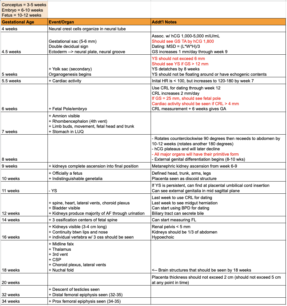
Hi Allison,
For the dates, my CI has this wheel she uses to check important measurements. I will bring it to class one day to show you all.
I was chatting with my techs today and they gave me some advice to get the LVOT and RVOT views. When you get the 4 chamber heart, angle the probe slightly towards the baby's head to get LVOT and then tilt towards the baby's right shoulder to get RVOT. The movements are very minuscule and be sure to keep in mind the baby's position when deciding which way to tilt your transducer. For instance, if the baby is in the cephalic position then the right shoulder would be downwards.
Hopefully this helps and you can try these tips at your site soon!
Good evening cuties,
Unfortunately for me I do not get much practice at all scanning OB so I have been using these discussion boards to learn from all of your experiences at your sites. Because of this, I have not been able to scan much at all with the heart and that is what I have been struggling with the most. I feel confident about the fetal circulation but when I see an image I get stuck. I try my best to just keep evaluating pictures and watching different videos (Thank you YouTube) to see different views but still find it difficult at times.
This a video that Dr. Wilson had shared that I think I have watched more than 5 times and it gives some great tips:
https://www.youtube.com/watch?v=Wreu8FkQRQE
If anyone has any other great resources or tips, please share.
Same here Raman,
I get a 1st trimester once in a blue moon and sometimes theres no baby in there so I'm basically just doing a pelvic exam, so sad. I was just saying the same thing in Leah's post about how I don't get to practice what we're learning and it's such a bummer. Hoping I get some exposure soon or at my next site! until then, keep the videos coming lol
Hi friends,
I'm really loving all the posts so far and you've all touched of things that I myself need extra time with and I'm definitely going to be studying up over the break. One thing I have a hard time with is memorizing fetal circulation. I drew it out but like Lania said in her post, my brain doesn't want to hold it to long term memory. I feel like maybe drawing it a few more times and watching some more videos may help but please let me know if anyone has a trick up their sleeve for this topic lol I found these helpful videos to review!
https://www.youtube.com/watch?v=wYNY7VpZIPY
https://www.youtube.com/watch?v=-IRkisEtzsk
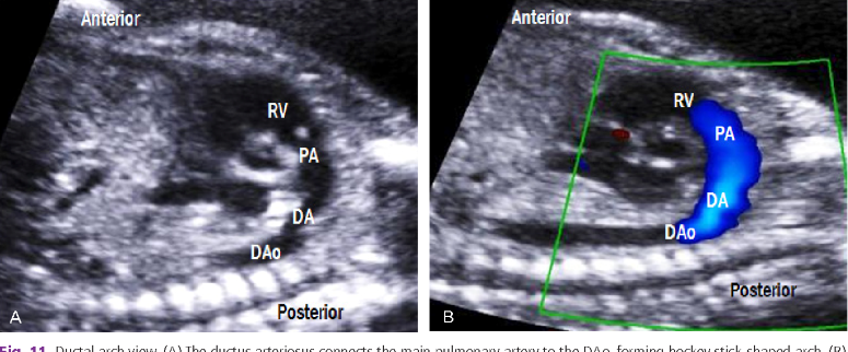
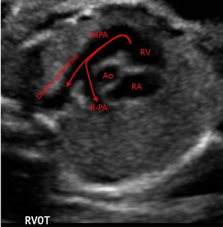
Wow, times flies! It is mind-boggling how much we've learned in this class alone. One thing I know we've talked about, but isn't sticking with me is short vs long axis RVOT. I know that they look different and present slightly different information, but which one is preferred (if any) and why? One of the techs said it depends on the site/rad's preference. Does anyone's site do both?
Hi Alexis!
I struggle with this too. I try to remember short axis RVOT by remembering it looks like a snail! I believe that the long axis RVOT can either be taken slightly above the 4 chamber view like the LVOT or it can also be taken in the 3 vessel view.
I think the preference of these types of RVOT views typically depends on the radiologist's preference. At my site the tech actually did both views because she wanted to show where the ductus arteriosus meets up with the aorta (long axis view) and the bifurcation of the pulmonary artery (short axis view).
I think Dr. Wilson also said that a lot of radiologists like the short axis RVOT view to prove that it does in fact bifurcate.

Hello everyone!
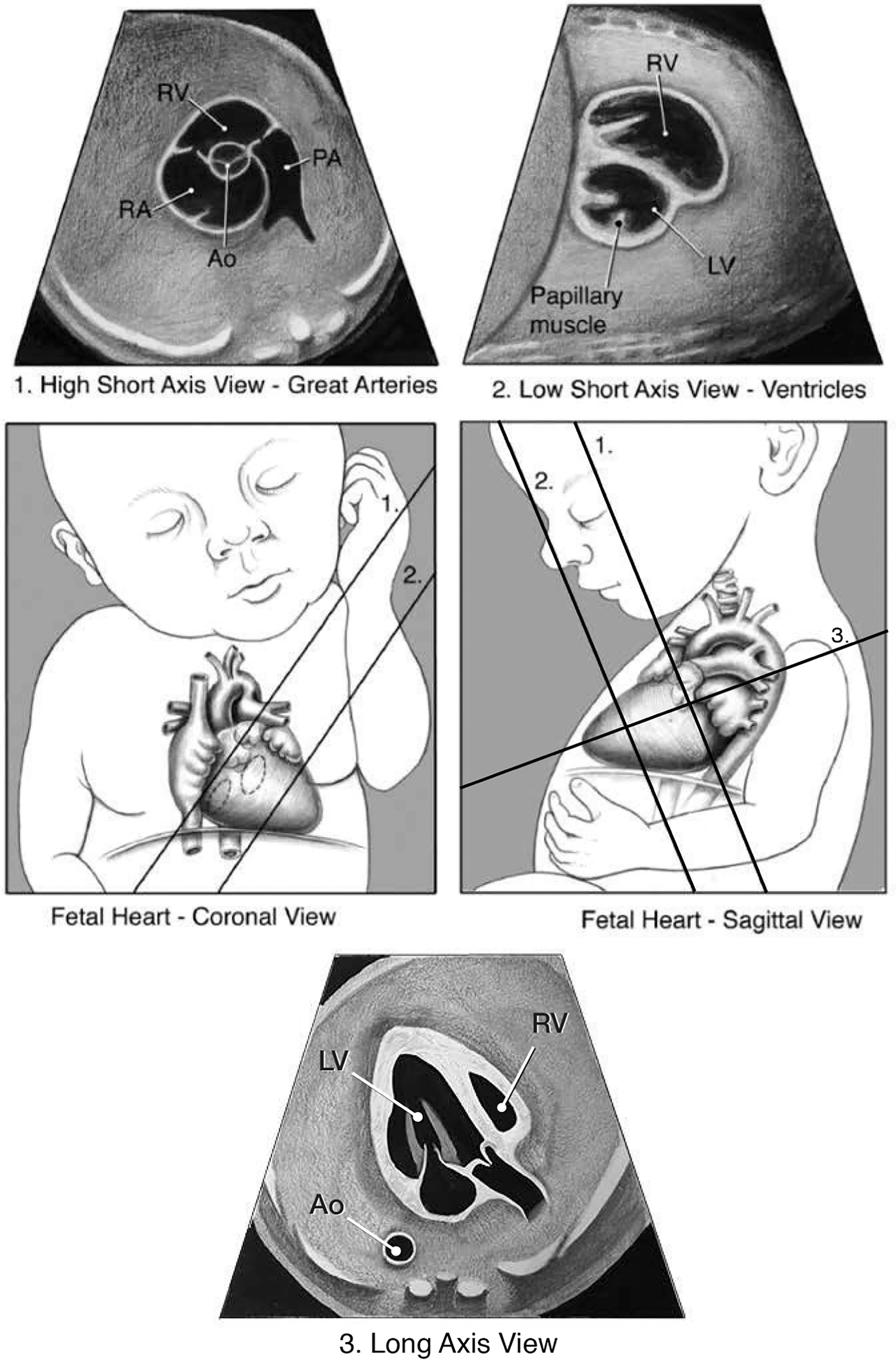
This class has gone by super fast and boy has it been challenging! There are many topics that I feel I need more time with including the fetal heart views and learning how to find them intentionally while scanning instead of by luck at this time, but a topic I feel is super important that I want to get down first is fetal lie. Beginning each OB exam I always spend time to try and figure out fetal lie, but it has been difficult to feel confident in telling my CI how I think the baby is positioned. My CI brought in a little doll for me to use that I bring every day to clinical's and it has been of great help. I think starting at the cervix and establishing if baby is positioned in a cephalic (head down), breech (bottom/feet down), or transverse position is very important to start off with. Next would be to scan in a transverse sweep from cervix where the baby's bottom or head is located and sweep superiorly until you reach the end of the baby so you're able to determine where the spine is at along with stomach/heart positioning.
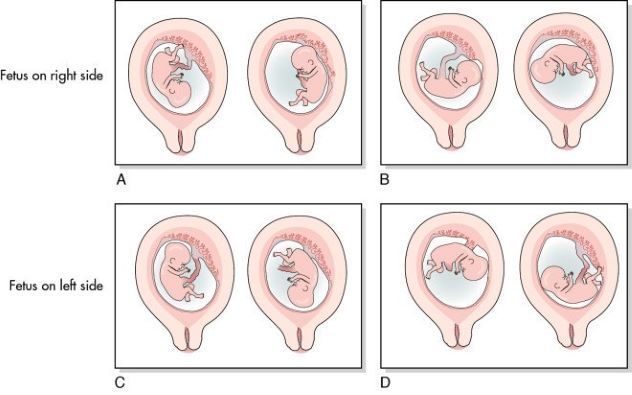
I found this diagram super helpful of the babies in both breech and cephalic and how the stomach/spine would appear on the monitor while in those positions. Also this YouTube video of fetal lie/determining situs is extremely informative. It is a but long but it helps a lot!!
https://www.youtube.com/watch?v=bXHM0t-kkrE

Hi Molly!
Fetal lie is a difficult one sometimes! I'm not sure if its at the very beginning of your protocol too, but I've found those initial rt to lt and inf to sup sweeps to be so essential for me determining lie right away! I had some trouble at the beginning because I would see, for example, feet at the cervix but the rest of the baby was lying sideways in the uterus so I would call it transverse. My CI had to tell me at least once "however the baby is lying more superior, if feet are at the cervix, they're breech". I had to look up transverse lie images this weekend too since we see that less and I can never confidently say transverse, so if we see abdomen presenting to the cervix or maybe upper extremities, they should be transverse.
You found some great images, so I think it's one of those things we just have to scan a few times and get more confident with to be sure!
Hi Lauren!
Thank you for sharing what your CI said about determining transverse vs breech. That is super helpful because I too have seen feet at the cervix and it seemed as though baby was lying across moms abdomen in a trans position, but it's nice to know it was considered breech. I definitely agree with you that time will make us feel more comfortable and knowledgeable with determining fetal lie along with the whole exam. I think we are all doing a great job so far!
Hi Molly,
I agree with you 100% fetal heart views and presentation are my kryptonite! I think one of the things that makes fetal heart so difficult is that a lot of the time is position dependent. While we may be able to see it, it doesnt always mean the baby is in the ideal position to capture an image (especially one a radiologist would approve). I always feel like I have "dumb luck" when it comes to heart, when I am not trying I can get a beautiful RVOT without even realizing or attempting to. I can't wait for the days when we have it down and we know exactly how to move our probe to get those desired views! When it comes to fetal presentation I had a small breakthrough today, I literally imagined I was the baby in the uterus and my mind was able to conceptualize how that position correlated with the image. I literally spent a decent amount of time talking to myself and squirming around in my chair before I felt confident with my answer lol. We just have to keep reminding ourselves that one day it will all make sense!!
Hey Chrishawna!
Cannot agree more with you that most of the time heart views just pop into view out of nowhere and I get lucky to view a beautiful LVOT OR RVOT. I feel like fetal presentation is preventing me from knowing how to angle my transducer perfectly to obtain those images intentionally. OB is so challenging and all of this material we've learned is great this quarter, but its one thing to learn how to get the heart view images, but when you're actually scanning a moving baby it's crazy challenging! Watching the techs at my site so effortlessly find each Ob image makes me hopeful for the future!
I am so so happy for you that you got to scan some OB today and you had a break through with fetal lie! I think it's an amazing idea to picture ourselves inside that uterus each scan we do until it becomes second nature to instantly be able to tell lie. Fingers crossed tomorrow we all get to practice OB!
Wow Molly I love these images, I've been trying to picture fetal position in my head because I don't see a lot of OB at my site. but today i got to observe a 3 trimester follow up! I was so excited. I wasn't able to scan her because she was a double study and she was already there for a while so the tech just did it real quick. but I learned a lot from it because they tech was walking me through it as she was scanning which was nice.
Anyways congrats on the LVOT girly you're doing amazing!!
The fetal heart circulation and identifying heart views is definitely something I am still working on. At my site we don’t really conduct any in depth OB studies, so I haven’t had a chance to put many of the scanning tips into practice. However all the tips everyone has shared have been really helpful as I try to figure it all out in my head during a scan. Although it’s so intimidating, I think for me sometimes what helps is just quizzing myself on ultrasound images, I wish there were more quizzes online so we could practice. I found this slideshow, it’s nice to quiz yourself on. Going over images many times and trying to recite it or draw it out by memory is always helpful as well! I think I'll be taking a small doll to clinic this week as well like Michelle suggested! (:
https://www.slideshare.net/orchard238/labelled-fetal-heart-protocol

Hi Zuly!
Fetal circulation can definitely be tricky. It helps me to first remember normal adult circulation and then add in the "short cuts" that fetal circulation can take.
I like to remember that there are 3 short cuts - ductus venosus to bypass liver and get into the IVC, foramen ovale to bypass the right ventricle and enter the left atrium, and the ductus arteriosus to bypass the lungs and get straight into the aorta.
Here's an image of normal adult circulation to help you compare the differences between the two! :)

Hey all,
I am still struggling on cardiac embryology. I am just not sure how in depth I need to know. This video (http://www.scielo.br/img/revistas/spmj/v137n5//1806-9460-spmj-137-05-391-gf2.png) has been the most helpful so far. I am just not sure if this amount of information is sufficient enough.
I think I have an understanding of what RVOT and LVOT look like but I think trying to understand what they mean. Is RVOT the outflow tract that is describing the pulmonary valve (where blood can the pulmonary artery or ductus arteriosus) and is LVOT the outflow tract describing the aortic valve (where blood travels through the aorta and goes to brain and body)?
I also have been struggling how to understand how the baby is laying in clinic. I do need to brush up on scanning planes because it plays an important role in this understanding.


sagittal

coronal (trans)
Hey Nailah!
I feel like we're all working on improving on the same topics, these are definitely the most challenging to nail down. I love that you mentioned going back to the basics, and revisiting the scan planes. I love this image Sue shared with us demonstrating uterine presentations transabdominally and how they will appear on EV. Scanning EV definitely flip flops my brain if the patient has a retroverted uterus!

Nailah & Zuly,
Both of you guys have shared great graphs that have definitely been a lowkey struggle for me. During clinicals, as I watch a OB scan, I watch where the baby is laying and investigate it's head, limbs-- the over all position and then "lay myself into the womb". I've honestly practiced this with a tiny doll that my CI had in his room. It's a lot of practice to figure the fetal lie. I take this challenge as a puzzle piece-- just got to take it one by one.
Thank you guys for sharing such great images again!
Howdy Nailah,
In order to determine fetal position I like to first check the cx area. Do I see a head, feet. or part of the torso? This will determine if baby is cephalic, breech, or transverse. Next, place the transducer at a 90 degree angle with the bed. Find the head and slide the probe from right to left. Do you see a face first or do you see the nose and lips come in after you've scanned through the head? This will determine if baby is facing right or left. This assumes baby is cephalic or breech. Adjust for transverse presentation as needed.
If you don't see baby's nose/lips this way, then baby may be spine up or spine down. Check by scanning at a 90 degree angle with the bed. Is the spine directly in line with the transducer beam? Is the spine closer to the footprint or further away? You may need to look transverse and long to double check.
Hope this helps,
Sarah
Throughout this amazing quarter with Dr. Wilson and Paris, I've struggled with understanding the heart views. There are time where I can pinpoint the structures of the descending AO, a lower case "r" that indicates the PA and know that it's RVOT or see LA, LV, and RV to know that it's the LVOT. But when I am observing some of the technicians at my site scan the segment of the heart, it looks so different. And that's where I get lost. I don't know if its because I'm over thinking this concept or just straight confused.
After some research, I found these fetal heart videos! Provided by Philips
https://www.youtube.com/watch?v=XTi_JtdnMJM
https://www.youtube.com/watch?v=jDl_XpGUJEU
https://www.youtube.com/watch?v=rW2r55AS3Xk&t=9s
https://www.youtube.com/watch?v=5arsJCjTqGg&t=1s
Sometimes, I wish there was a TED talk about what ultrasound students go through. This quarter wasn't easy, but then again "Nothing good is ever easy". During break, I still plan on drilling and keeping everything stuck in my head. I would hope that all these concepts become second nature to us someday.
I'm super excited to see how these discussion post plays out! Team work makes the dream work.
Hi Monica,
I totally agree with you and struggle with heart views as well. :( I noticed when I see the views on paper I am able to somewhat understand it but when I watch a tech scan, I get so confused and am not able to follow along. I remember that Sue was saying as a tech our eyes are always on the screen and she does not pay much attention to what her hand is doing. Since you scan OB at your site, do you try to angle to get your different heart views or do you just get a 4 chamber and follow the screen to see what you can get? I feel a lot of sites say to angle a certain way but in reality when working with a moving baby (especially a hyper one) that may not be the most practical approach.
Hola Raman,
Honestly, I will try to get a 4 chamber view and try to complete the rest... but these babies are tough! They move around like crazy and I don't want to spent 5- 10 mins chasing them down. I think if I'm able to manipulate my transducer within smaller movements, I could accomplish these images. But I'm not close to scanning OB like a pro.
Hey Monica!
Heart views is something I can understand visually, but to actually get them sonographically is something entirely different!
Do you get to scan at all, or do you have to just observe for now? I feel like getting hands on experience is really key to getting these different views!
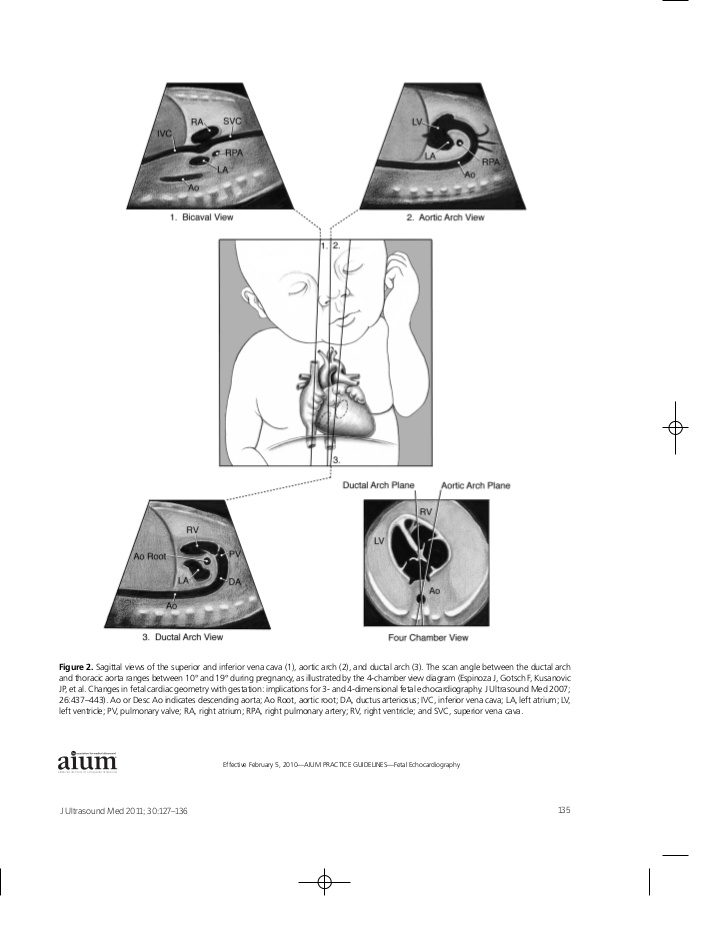
I've compiled some different scan planes for fetal heart that may help us.

Hi Monica!
I'm with you love! Some days I get them and some days I don't get it at all. I've been watching YouTube videos to try to help which I find helps me recognize what each view should look like but it doesn't necessarily help me find the actual heart views. My CI told me that one day it just clicks and the best way to learn it is just to keep scanning it and it eventually becomes like muscle memory! So we will get there one day! But like you said, nothing good is ever easy, but we got this!
Hi ladies!
Looking back on this class, we learned a true wealth of knowledge in just one quarter! I really thank Dr Wilson and her way of teaching, which was so foreign to us in the beginning but ended up working so well. I feel like we truly absorbed a lot of what we learned.
One of things I still struggle with is remembering fetal growth measurement is not the same as checking fetal age. I got that mixed up on today's quiz... when I saw growth I was thinking age-wise and I picked the wrong answer. I think a few of us also struggle on the terminology and reading questions too quickly.
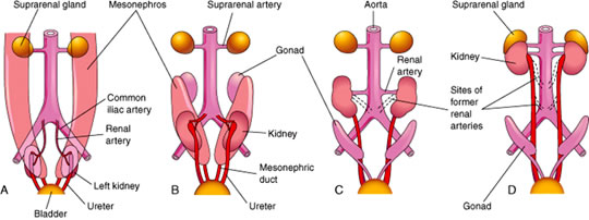
Another would be renal embryology and normal measurements. I was looking into pictures and saw a few that hopefully can help others who need it!


Hi Karen,
Thank you so much for sharing such an informational image! I am going to print it and put it in binder. Very simple and easy to follow.
Why do you think knowing embryology is so important for OB? I was asking some of the techs at my site and they did not know some of their embryology lol
Hi Raman!
I think it's important to know embryology for OB because we are essentially watching the little organs grow as we scan! Throughout a person's pregnancy, I mean, not right in front of our eyes :P
It really helps to know how and why things go wrong, and knowing how things are formed is a big variable in the equation.
I hope we remember as much as we can going into our careers!
Hi Karen! I feel you on reading questions too fast. It's always a bummer when I re-read it after and I know the answer >.<
I made this chart to help me organize the anatomy scan measurements. I think I may want to add renal pelvis measurements though, just in case there is questionable dilation. If you can think of any other ones I missed, please let me know!
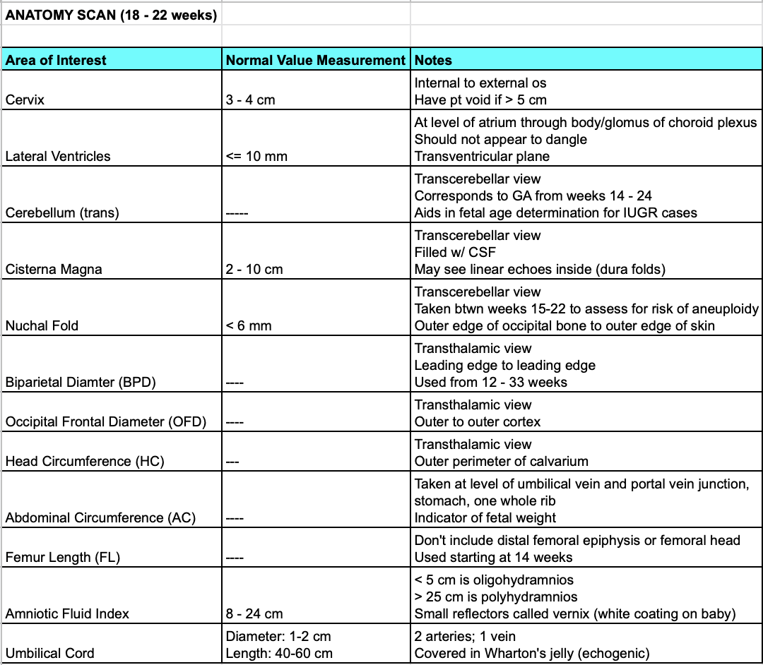
Howdy everyone,
My main confusion is seeing the cervix trans abdominal. I can normally see where it is, but I'm not always sure where to put calipers.
Additionally, if anyone has any tips or tricks on remembering what happens at what week I'd be grateful! I think I'm going to make a document with all the dates and milestones listed.
Sarah
Hi Sarah!
Cervix is always a struggle for me as well... Usually it's not as clear as this but I found a nice example for measurement!
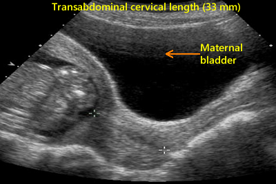 We may just need to angle better to fully utilize bladder as a window.. but I think sometimes it's just hard to really see. I'm guessing this is where years of experience comes in, haha.
As far as development, I found this great chart! It includes time where baby is most at risk for abnormalities, which I thought was really interesting. I'll probably be referring back to this if/when the time comes for me to have a bun in the oven!
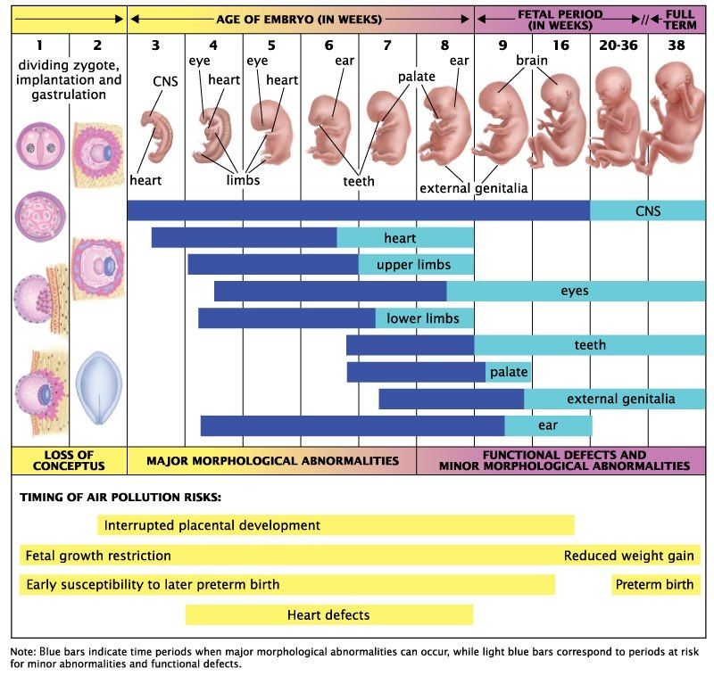
Hi Sarah!
I totally understand where you are coming from. I struggle with this from time to time. The best advice that my CI gave me was to just get as much exposure from OB. There's a lot that can change the appearance of the cervix/ os and placenta images (Braxton Hicks contractions). The cervix could look different due to the mother's bladder or the fetal head giving us some side wall shadowing! It's important to just take a deep breath and remember the landmarks that Karen's image shares above. Overall, I think you're doing great!
Hey Sarah,
The trans cervix is hard for me too especially if the woman is not pregnant. I think its more of a judgement call but it kind of seems like early on in the cycle it is harder to tell but once the endometrium gets thicker you can kind of see it thinner in the cervix area

This image kind of shows it best! See where the endometrium gets thinner and the cervix tissue also is a bit more hyperechoic.
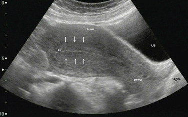
Cervix measurement is definitely a tough one both transabdominally and on EV!
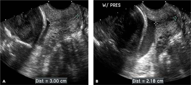
Today one of my techs and my CI had a discussion about this very topic, in regards to a scan I observed on a 3rd trimester OB patient. She had a cervical length order, and on EV it was immediately evident she had cervical funneling. No matter where she placed them, it was on the shorter end, but depending on where she put them, it would make a difference between lower limits and definite short cervix.
My CI said we should have applied pressure with a hand, on the pelvic region, while performing the EV scan, to see how it affected the cervix. I'm not 100% sure what information this would give us, but I thought it was interesting. In the end my CI said the cervix measurement, particularly the caliper placed on the external os, is somewhat subjective, but to be mindful not to extend it into the vaginal area.
https://radiologykey.com/uterus-and-cervix/
- The length and shape of the cervix may change during the course of the sonogram. This can occur spontaneously (Figure 17.1.4) or may be elicited by manual pressure on the uterine fundus (Figure 17.1.5). In either case, the likelihood of preterm delivery correlates with the shortest length of the cervix during the sonogram.
- Cervix dilating with fundal pressure .A: Sagittal transvaginal view of the cervix demonstrates a normal appearing cervix measuring 3.00 cm in length. B: With manual pressure on the uterine fundus (W/ PRES), the cervix shortened to 2.18 cm in length.
Hi Zuly!
Thank you for sharing this information on the maternal cervix! I have also seen my techs place a hand onto of the maternal pelvic region and try to gently push baby's head away from the cervix to get an accurate measurement while performing TV.
I have also seen funneling with a shortened cervix like the example here in B. Very important to acknowledge the differences here to insure we never get a wrong measurement. Always making sure the maternal bladder is empty and the baby's head is away from the cervix during the Transvaginal study for an accurate measurement is key.
Thank you for sharing!
Hi Sarah,
I always remember that the rhombencephalon is seen around the 7th week.
Rhomben-SEVEN-lon!
Hi Sarah!
I too struggled with seeing cervix and still do a bit, but my CI told me a trick to look for the shape of a U. It can be hypoechoic or a hyperechoi or even homogenous which I think is what trips me up because everyone's cervix can look different, but once she told me that I started looking for the U shape and found it a bit easier to spot. I hope this helps a bit! :)
Good morning everyone,
Thank you all for sharing such great and amazing tips with each other! Team work makes the dream work! Remember this course is available to look back through so we can go back and rewatch a lot of the videos and images that were shared. I think one of the biggest struggles for all of us is embryology and the heart. As many of you mentioned, knowing normal circulation and then comparing it with fetal circulation is a nice way to remember the different pathways. Also, be sure to always keeps in mind baby's position while scanning different anatomy so do not be hesitant to do a quick transverse sweep whenever you feel the baby has moved and you need to establish its position again. Being able to correctly orient ourselves and the image is crucial in our career so reviewing the basic planes and having a quick cheat sheet is always helpful. We need to all have a little doll party next quarter in lab where we can practice fetal orientation and it will help in letting us analyze different windows with our transducers to see the baby's anatomy much better. As far as embryology goes, I think with repeated repetition and rewatching the videos again and again, we will be able to lock it in. With time and practice, it will all become second nature to us and we are gonna think how silly we were to think all this was so difficult. I mean even now, just look at our progress from the beginning of this quarter to now the end. We will take all that we have learned in this class and use it to help the lives of babies and their families.
Thank you Dr. Wilson and Paris for teaching us so well! You guys are great teachers and will be missed next quarter. <3

