What are the earliest sonographic signs of pregnancy and which ones are definitively reliable for a diagnosis by the healthcare provider (OB/Rad/NP/ED Dr./ RN Midwife) that this patient is pregnant?
Week 2 DB: Conception/Fertilization and 1st Trimester Scanning
Sonographic Documentation
Re: Sonographic Documentation
Hi everyone, this week I will be the moderator for this discussion board, I'm excited to see what all your beautiful minds have to say about the earliest signs of pregnancy with sonography. Keep in mind not to just post a list of things, explain how you came up with that and what role does it have in pregnancy? Think of some possible things that can go wrong if we have one sign but not another, what have we learned so far that can effect a viable pregnancy? What can be mistaken for a pregnancy and what part of an early pregnancy should not be mistaken as pathology? Be creative but on topic with your responses.
Re: Sonographic Documentation
As we discussed today in lecture, the first thing we might see is the "intradecidual sign", which is essentially a tiny bubble that indicates some sort of pregnancy reaction is happening. However this is not super reliable. It does nothing to signify viability or if the pregnancy is even intrauterine.
Further research also brought me to "decidual cyst" and "pseudogestational sac". I found this article on Radiopaedia that explained pseudogestational sac and listed decidual cyst as a differential. Can anyone differentiate these? Or are they the same thing? It was a little unclear...
The gestational sac, which is the chorionic cavity, is usually the most notable first sign of a pregnancy with the "double decidual sac sign". However until there is a confirmed "healthy" yolk sac (usually see around 5-5.5 weeks), a healthcare provider likely wouldn't call it as pregnancy.

I'm really curious to hear about experiences of those who are in clinicals and what they have seen for OB scans!
Re: Sonographic Documentation
Hi Leah!
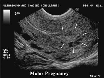
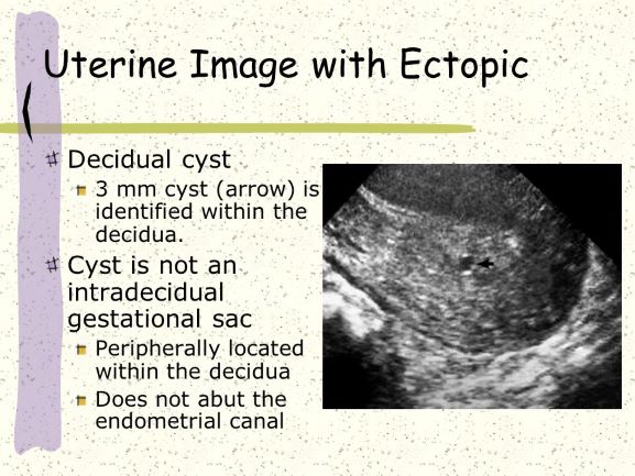
You had me curious about the differences between pseudogestational sacs and decidual cysts. I kinda ended up, down a rabbit hole in my research, but can say I think I may understand the differences. I found a really great article that is pretty short and to the point, but actually covers both topics (I love it when that happens)! From my understanding a pseudogestational sac is exactly like it sounds. Something that may sonographically mimic a gestational sac but is not one. Some articles I read through say to look for the double decidual sac sign around the gestational sac to see if it's real or actually pseudo, while other articles say not to rely too heavily on that sign as it may not always been seen on every sonogram. A decidual cyst on the other hand can occur as part of a an intrauterine or extrauterine pregnancy, and in women who are not currently pregnant (although this seems to be less common). Basically, as the decibel cells deteriorate, they can fill with fluid and become cystic, but are not of concern if they remain less than 3mm! I have attached the article so you can take a look, it also has some great sonographic images: Ectopic Pregnancy
Re: Sonographic Documentation
Hi Leah and Chrishawna,
I love the research going on! I hope this help you Leah. If there does happen to be a gestational sac and it grows to be around 12mm and theres no yolk sac in sight, what would this mean for the pregnancy?
Re: Sonographic Documentation
Great question Hailey!
If there is a gestational sac that reaches 12mm and there is no visualized yolk sac, this would indicate a failed pregnancy. According to my class notes, the gestational sac usually reaches 12mm around the 6th week and the yolk sac appears in the 5th week, so there is a ~7 day window for the yolk sac to appear before there is cause for concern.

Re: Sonographic Documentation
Hey Ms. Porter
Thank you for sharing that article.I found it really helpful to have some visuals of ectopic pregnancy! It can get tricky especially when the ectopic is just outside the uterus. I also found that gestational age to hCG levels comparison chart extremely helpful. Great resource Chrishanwa!
Best,
Nailah
Re: Sonographic Documentation
Hi Leah,
I realized our responses are similar! I also mentioned the pseudosac. I definitely believe that a truly viable pregnancy should be called when you can see the yolk sac, a sign that something is alive and growing!
I couldn't for the life of me find a picture of a decidual cyst BUT I did fond something to support our argument:
Endometrial cysts that could kind of look like gestational sac.
Re: Sonographic Documentation
Hi Leah! Great images and response. Since we've learned about the proper identifiers for early pregnancy, it actually made me think about on my first day of clinical we did and exam on a patient for early pregnancy and I want to say she was around 6 weeks and there was no sign of a yolk sac yet. Of course then I wasn't really aware of what I was looking at but now looking back I believe that was abnormal, and plus that patient had a history of miscarriages which was super sad, but just knowing now what is normal and abnormal now definitely opened my eyes.
Re: Sonographic Documentation
The earliest sonographic sign that could suggest pregnancy would be the visualization of the gestational sac around the 4th week. For some, this may be a clue that the pregnancy is forming, but would not be definitive proof until a week later when the secondary yolk sac is seen within the gestational sac. I recall Dr. Wilson saying she would simply describe the sonographic appearance of the gestational sac and not refer to it as an intrauterine pregnancy until after the secondary yolk sac has formed. By week 6 the fetal pole should be noted with the presence of cardiac activity.
Another sonographic landmark to indicate gestation is by locating the double decidual sac sign which is created by the presence of the decidua capsularis and decidua parietalis. I am also curious to know if a corpus luteum cyst could be considered a sonographic sign since it is typically present throughout the first trimester if it were accompanied by another sign like the gestational sac. I know these two can often be mistaken for one another, but if both are present maybe it could be a sign?
Although there are many potential sonographic indications of pregnancy the most reliable in my opinion would be around week 5 to 5.5 when you should be able to document a gestational sac along with an enclosed secondary yolk sac! Please let me know your opinions on the corpus luteum, I know they can occur outside of pregnancy, but do you think they could be significantly if a gestational sac was also present?
Re: Sonographic Documentation
I find myself spread out thin when our textbooks goes over the same concept, yet the numbers somehow are always off. It's interesting that you mention that the presence of gestational sac would be 4th week. Questioning your corpus luteum cyst/ double decidual sac sign-- that would be an interesting presence! I found an image that shows the two together (shared below). I also agree with you that the best sonographic appearance to confirm a pregnancy, sonographically, is week 5: the presence of the gestational sac within the chorionic cavity.

Also really I appreciate you restating Dr. Wilson's comments upon this subject! I was missing some notes. You're the best.
Re: Sonographic Documentation
Hi Monica,
I love the image you shared, how cool for the sonographer to be able to capture two signs of pregnancy within one image! Going back to what you said about the timeline for events being off, depending on the source they come from, is something I agree with. As a student its hard when you're just trying to lock something in, but different websites have opposing information. From what I understand we can see the pregnancy at different weeks depending on what method of imaging we are using. Endovaginal imaging seems to be one week ahead of the time that we can see the same thing on transabdominal. For example, if we were to scan a 4-4.5 week pregnancy using the transabdominal approach we may not be able to see the gestational sac. If you then use endovaginal you may be able to clearly delineate a gestational sac on that same patient within the same appointment. I think this is why there might be a slight discrepancy on the weeks and also why many technologists will use both approaches! Here is a short study done to show the differences between the use of EV and TA in the first trimester: https://pubmed.ncbi.nlm.nih.gov/21966152/ I also included a great image from Dr. Wilsons presentation that shows the time variance between EV and TA!
Re: Sonographic Documentation
HI Chrishawna! I love how you emphasized what Dr. WIlson said about not referring to the preganancy as an intrauterine pregnancy until the secondary yolk sac is formed. Even if the gestational sac forms, it could get up to 12mm and still not contain a secondary yolk sac which would mean the pregnancy failed. So I think it's super important to keep that in mind and not jump the gun at saying someone is having a successful pregnancy just because a gestational sac is identified.
Re: Sonographic Documentation
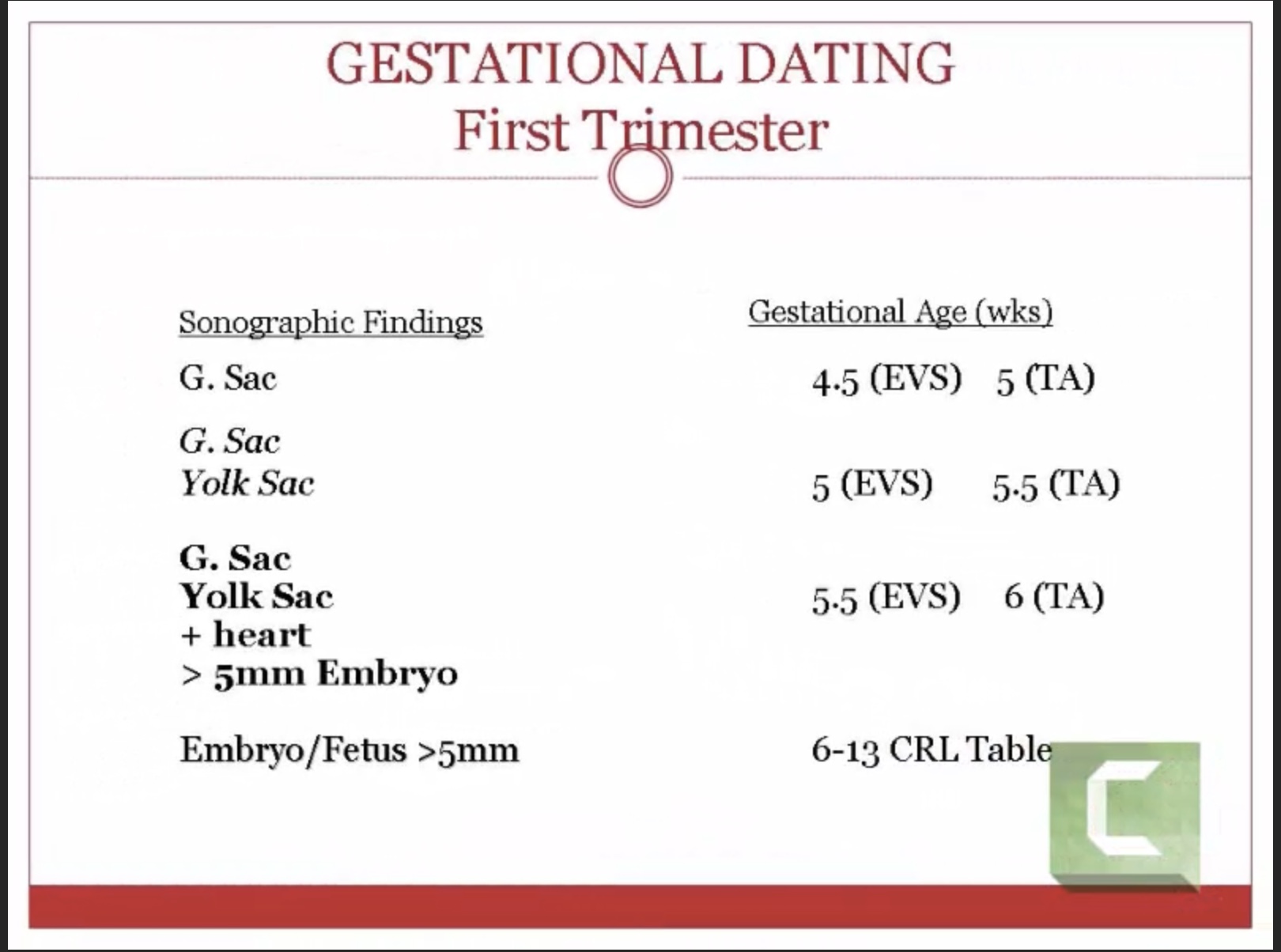
We may be able to detect pregnancy with sonography, at 11 days after conception. Sonographically, we would find the intradecidual sign, a round fluid collection within an echogenic area, beside the endometrial cavity. I say 'may be' because, if I am correct, this sonographic appearance is also the same as an ectopic pregnancy. Although, the most reliable sign for a diagnosis by the healthcare provider that this patient is pregnant is viewing the gestational sac at 5 weeks or with the gestational sac and yolk sac at 5.5 weeks.
Also, the presence of the gestational sac will be correlated with HCG levels, a hormone produced by the placenta that also signifies a positive pregnancy.

Re: Sonographic Documentation
The earliest signs of pregnancy is first the formation of the gestational sac which is seen at 4 weeks,grows at about 1mm a day until the 9th gestational week, and has a double decidual sac sign around it. Then the yolk sac forms at the 5th week, which should be measured between 2-5mm and not to exceed 6mm if there are not issues such as a nonviable pregnancy. The fetal pole then forms at 6 weeks, which is where a fetal cardiac movement can be visualized and measured. In some normal pregnancies the fetal cardiac heart beat is not seen until the fetal pole measures 4mm in length. The gestational sac with a double decidual sac sign is the most reliable for a healthcare provider to diagnose pregnancy.
Here's an ultrasound image found online that demonstrates the gestational sac with the yolk sac and fetal pole within it. Does the gestational sac look as if its round and an appropriate shape for a viable pregnancy? I remember from Dr. Wilson's lecture that if the gestational sac started to look as if it were compressing then it may not be a good sign for the pregnancy. This gestational sac doesn't look very compressed to me, but may be something to still document since it looks as if its starting to compress near the fetal pole?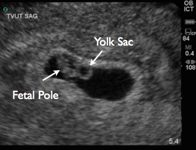
Re: Sonographic Documentation
Hi Molly,
I like that you included the timeline in your response, also your question is very valid, this doesn't seem too alarming to me but I did find a case online that said a patient came in for bleeding, they gave her an US and her GS was very compressed and no fetal pole was found. it ended up being a uterine rupture. Is there much of a difference in these pics? how does this explain the importance of history?

Re: Sonographic Documentation
Hi Hailey!
Very interesting photo you found! Compression of the gestational sac is very interesting to me and will be useful to know this information for our career. Our photos do look similar except yours does show that the gestational sac is much more compressed and the fetal pole is not evident like you mentioned. I think patient history is extremely important because these are symptoms and instances that can lead to an uterine rupture: severe abdominal pain, hemoperitoneum, a history of a prior cesarean section, dilation and curettage, or even uterine surgery. A good grasp on patient history lets us know a lot of valuable information to use in order to connect the puzzle pieces while scanning.
Thank you for your question!
-Molly
Re: Sonographic Documentation
Amazing! Today in clinic my CI asked me if I had a 2-5 year old come in with diffuse abdominal pain, what would I think about right away! I have to get better at thinking about these things because I know they will come my way very soon, that's why I'm loving our critical thinking class because it's making me think! I think theres also another aspect to this as well, just because they have pain in an area it doesn't guarantee that there will be pathology. The human body is so complex and sometimes imaging can't pick up the problem.
Re: Sonographic Documentation
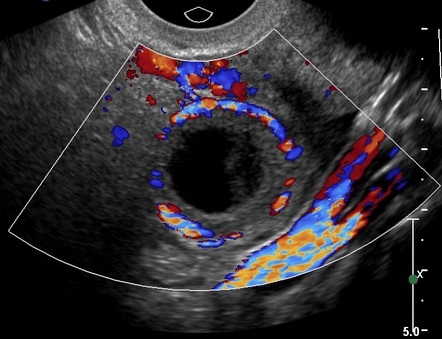
Some early sonographic signs of pregnancy could be a gestational sac that has a double decidual sign. Or even a cyst like sac and a positive HCG test could indicate there is a pregnancy but could possibly be too early to tell or is ectopic. However, more reliable signs would be the visualization of a yolk sack within a gestational sac with a double decidual sign, a fetal pole and even cardiac activity. I believe also that a corpus luteal cyst with the above findings may also correlate to a pregnancy. Here are some examples
Sac or Cyst within the Uterus & Corpus Luteal Cyst

Gestational Sac with double decidual sign, Yolk sac

Gestational Sac with double decidual sign & Fetal Pole
Re: Sonographic Documentation
Hi Allison!
I love that you mentioned how a cyst like sac may be seen in the uterus, but that this doesn't mean there is an intrauterine pregnancy. This is something that really stood out to me during lecture yesterday because I know as sonographers we may be excited to see what looks like a gestational sac, however a yolk sac must also be seen to rule out the possibility of an ectopic pregnancy. I remember Dr. Wilson explaining how these gestational sac impostors can be formed with an ectopic pregnancy because the uterus still responds to hormonal changes and its cells will continue to prepare as if it is an intrauterine pregnancy. It's fascinating to me how as soon as the body starts receiving signals that it is pregnant the uterus begins developing a nice home for the embryo!
I added a little graphic to my original post which talks about the decidual cysts seen with ectopic pregnancies and how they can mimic gestational sacs. Feel free to check it out if you want a little more information! :)
Re: Sonographic Documentation
Hi Allison and Brittany,
Love the convo here. Quick question, Brittany you mention hormonal changes. lets say the mothers progesterone levels drop in early pregnancy, what do you guys think would happen next?
Re: Sonographic Documentation
Hi Hailey!
Progesterone is a very important hormone with regards to building up the endometrial lining and maintaining it. This hormone typically has high levels during the luteal phase of a woman's cycle, so it can prepare the uterus for a pregnancy. If these levels are low, the endometrial lining will not build up and instead may actually shed. This is what happens during menses; when estrogen and progesterone drop really low the lining is sloughed off. If this level drops during pregnancy it is detrimental as it can cause a spontaneous abortion.
Re: Sonographic Documentation
Hi Allison! I love the photos you added, super helpful. I agree with everything you pointed out about being an indicator of early pregnancy. However, I've always been curious if a corpus luteum cyst would be seen in EVERY pregnancy? And if it's not seen could that be a bad indication for the fetus and the health of the pregnancy. I tried to look it up but didn't really find anything. Do you know or any of you other lovely ladies???
Re: Sonographic Documentation
Hi Lania!
I had the same thoughts as you regarding the corpus luteum, since it has such a huge role in secreting progesterone to keep the endometrium thick until the placenta can take over I think it has to be present in every pregnancy but I am wondering if it is not always sonographically seen? Here is a cool article I stumbled upon that is all about the corpus luteum in the first trimester mentioning it was detected in 95-98% of women between 5-9 weeks!
https://www.fertstert.org/article/S0015-0282(07)03643-6/pdf
Re: Sonographic Documentation
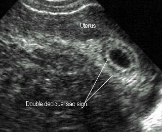
One of the earliest signs we might see on an ultrasound is the double decidual sac sign. This sign appears as two echogenic rings and is from the decidua capsularis and decidua parietalis. This shows that the uterus has begun preparing for pregnancy. However, this sign alone is not definitive of an intrauterine pregnancy.
A gestational sac is also an early sign of a pregnancy that may be seen around 4 weeks gestational age. This appears as an anechoic structure in the uterus. However, as we discussed in class, sometimes the intradecidual sign that may be seen is actually not the gestational sac. There may appear to be an anechoic "bubble" in the endometrium, which may mimic a gestational sac, but it actually isn't. With an ectopic pregnancy, the uterus may still respond as if it were an intrauterine pregnancy, and thus it still grows and develops in response to the hormonal changes. This is why it is very important not to call the anechoic structure a gestational sac or intrauterine pregnancy until a yolk sac is seen.
Perhaps the most important early sonographic sign for a pregnancy is the visualization of the yolk sac. The yolk sac appears inside the gestational sac as an anechoic structure with an echogenic outside ring. This structure will provide nutrients for the developing embryo and is a sign of a definitive pregnancy.
Re: Sonographic Documentation
I love how you described exactly why the first two possible signs may not indicate a true IUP every time.
Ectopic pregnancies are so interesting to me because of how the uterus and body still prepare themselves and show the same signs as they would if there was an intrauterine pregnancy. It really shows how there are multiple pieces that must come together for it to be successful.
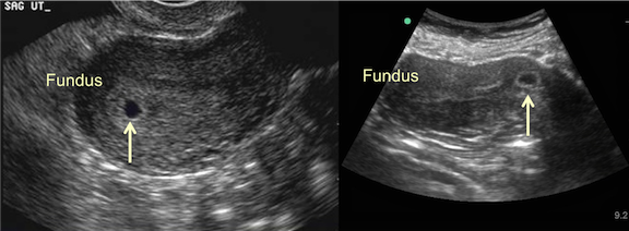
I think it is important to note that we should be seeing these signs, including the gestational sac and round, echogenic yolk sac, in the fundal area of the uterus. We may see these appear properly, but if they are too low and in the cervix, the pregnancy has a higher risk of miscarry.
Re: Sonographic Documentation
Hi Lauren!
You bring up a good point about how the location of the gestational sac is just as important as making sure there is one! I did a little research and learned that there's something called a cervical pregnancy which can happen when the gestational sac is formed too low in the uterus. Most of these unfortunately undergo spontaneous abortion.
Here's an image to show the different locations! The image on the right is a gestational sac near the cervix.
Re: Sonographic Documentation
Early sonographic signs of pregnancy include the intradecidual sac sign, double decidual sac sign, and visualization of the yolk sac within the gestational sac.
The intradecidual sac sign appears at around 4 weeks, and looks like a small anechoic fluid-filled “cyst” within the uterus, which represents the gestational sac
(which I believe is the sonographic term used to describe the chorion; but correct me if I’m wrong please!). It’s important to note that this anechoic structure should not be called a gestational sac until a yolk sac is seen.
The double decidual sac sign is formed by the decidua capsularis and decidua parietalis which appear as two echogenic rings surrounding the gestational sac.
Visualization of the yolk sac is another early sonographic sign of pregnancy, and its visualization is important in being able to identify a gestational sac with more certainly, and in determining an IUP.
I found it very helpful that some sources referred to the visualization of the fetal pole and yolk sac, as the “diamond ring sign, here is an image in case it helps anyone remember!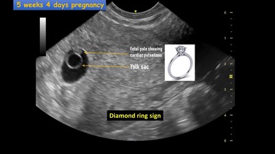
Re: Sonographic Documentation
Hi Zuly,
I never noticed that the fetal pole and the yolk sac actually do look a diamond ring! Thank you for sharing that. At first, I too was confused about chorion and gestational sac but after some research I have come to the conclusion that they are the same thing. Here's where I found my information at: https://www.glowm.com/pdf/Ultrasound_in_obstetrics_and_gynecology-chapter4.pdf
Re: Sonographic Documentation
I love the picture you added of how it really does resemble a ring!
I found the video that Raman posted in her initial response to be a helpful reminder too that the sonographer has to be careful not to document any yolk sac seen as a successful IUP, unless it shows a round appearance with good measurements of the gestational sac and decidual sign in place as well. I also remember Paris adding that a highly echogenic yolk sac (pictured) may be calcified, which is also a bad sign for the pregnancy.

Re: Sonographic Documentation
Great example of an abnormal yolk sac! I would also venture that this is an abnormally-shaped gestational sac. According to my notes, the gestational sac, much like the yolk sac, should have a rounded (or oval) appearance.
Below is a video and some images that indicate an abnormal gestational sac. This further supports that healthcare practitioners should not call it as a viable pregnancy without a yolk sac.
Empty gestational sac at 6wk2days: https://www.youtube.com/watch?v=YrXB4eMuMGI
According to this video, an empty gestational sac can also be referred to as a blighted ovum or anembryonic pregnancy, so it appears those 3 terms can be used synonymously. Good to know!
Re: Sonographic Documentation
Hi Leah!
Great examples of abnormally shaped gestational sacs. Today in clinic I had an interesting/sad OB case. The gestational sac was oddly shaped and it sort of tapered at the ends. Additionally there looked to be 1-2 or 3 fetal poles, with at least 2 cardiac pulses visible. The areas of the fetal poles were clustered sort of closely together and were very difficult to distinguish with certainty.
Some of the techs were discussing the case and were going back and forth on how many fetal poles they thought there may be, but in the end they concluded that regardless of the number, the pregnancy was "incompatible" (with life) because of the gestational sac shape Just goes to show how important it is for us to be able to pick up on and document such findings!
Re: Sonographic Documentation
Hey Zuly,
Thank you for sharing this sonographic sign! It is very helpful to know what you are looking for when things are normal. After doing some image research I did see that sometimes the "ring"/ yolk sac and fetal pole can be seen in the same image but sometimes, just depending on the position of the fetus and yolk sac you will see one then have to move or angle the probe again to see the fetal pole. Also I found a great image showing the double decidual sign as two echogenic rings. I sometimes have a hard time seeing the double decidual sign. To me it seems like one big hyperechoic band around the gestational sac. I believe also, and correct me if I am wrong, that the decidua basalis is identified as the part of the hyperechoic ring where the yolk sac and embryo are attached to the endometrium?
Re: Sonographic Documentation
Hi Zuly I liked your visual but was wondering when the yolk stalk would be seen and for how long would it be seen for. I don't see it in many pictures from around 4-5 weeks but I see them in others. The picture labeled it as the fetal pole which I understand is where the fetal pole grows from. Are these the same thing?
Re: Sonographic Documentation
Hi Candee!
Great question! I am not really sure when/for how long the yolk stalk would be seen, but while looking into it, I came across the "yolk stalk sign" which is described as a sonographic feature that suggests a non viable pregnancy. The embryo is visualized adjacent to the yolk sac in early pregnancy, and later, the yolk sac stalk develops. The yolk stalk sign is an abnormal separation of the embryo from the yolk sac, specifically when the CRL is 5 mm or less without a visible heart beat.
Link to the image/information source:
https://www.ncbi.nlm.nih.gov/pmc/articles/PMC5847503/
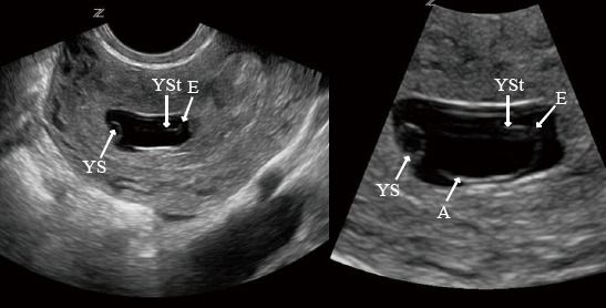
Image description: Transvaginal ultrasound image demonstrating yolk stalk (YSt), embryo (E), yolk sac (YS) and amnion (A). The yolk stalk allows marked separation between the small embryo and yolk sac.
Re: Sonographic Documentation
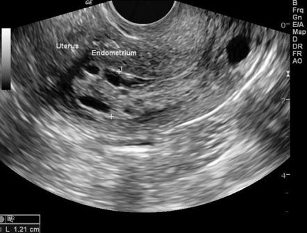
The earliest sign of a pregnancy is a gestational sac and that can be seen around week 4 of a pregnancy. A gestational sac will appear as an anechoic ovoid or circular shaped area in the uterus. However, one should keep in mind that one cannot definitively say a patient is pregnant until a yolk sac is visualized within the gestational sac. A cyst can also be mistaken for a gestational sac and it is common for the endometrium to thicken when there is an ectopic pregnancy. If a patient's HCG levels are high or they are having pregnancy symptoms but no intrauterine pregnancy is seen then we should look thoroughly to make sure there is no ectopic pregnancy. Our job as sonographers is to try our best to find any abnormalities so that the patient and baby can be safe. When looking for a pregnancy, we must first complete a trans abdominal scan to get a good field of view and trans vaginal to get more details. Thoroughly examining the adnexa is very important for an ectopic pregnancy. Also, if an intrauterine pregnancy is found then it should be in the fundus area, ovoid shape, yolk sac should be fairly circular and decidua should be thick surrounding the sac. As we can see in the picture below, there is a anechoic gestational sac with a yolk sac inside and a thick layer of endometrium surrounding the sacs.

Here is a very helpful video that I found for first trimester pregnancy: https://www.youtube.com/watch?v=4X6rcJwIFZg
Re: Sonographic Documentation
Hi Raman!
You make an excellent point about making sure we always scan the adnexa in pregnant patients! I remember last quarter, when we were learning about ovarian masses, how sometimes these masses can secrete hCG and make the woman think she is pregnant! I believe one of the masses that may secrete hCG is a teratoma. It's definitely important to look for these tumors to evaluate whether the patient is actually pregnant, either with an intrauterine pregnancy or an ectopic one, or if they have a mass that will need medical attention.
Re: Sonographic Documentation
Hey Raman,
I appreciate you noting the possible things that could go wrong and what we should be looking for if we do not see the typical signs of a pregnancy. I found this amazing website that goes into detail as to what are typical signs of pregnancy staring at 4 weeks. It also shows a lot of the possible abnormal findings. For instance, the ones we highlighted in class could be no yolk sac seen in a gestational sac that measures >12mm (image 1). Another could be a collapsed gestational sac which could mean fetal demise( image 2) and lastly this website says a gestational sac greater than 18mm without a fetal pole (image 3). I researched a bit more and I found that other resources claims 16-24mm without a embryo could also mean pregnancy failure.


https://www.acep.org/sonoguide/obgyn.html
Re: Sonographic Documentation
Hi Raman,
We are thinking along the same lines! I couldn't say that it is a positive pregnancy for certain until I see that yolk sac. It really could be anything else. This DB reminds me of a patient I scanned today that was positive for pregnancy, checking for ectopic, but at a little over 5 weeks there was no yolk sac yet. We were both just so relieved that it wasn't ectopic that I didn't think about anything else and I congratulated her on being pregnant. Now I am wondering-- what if it was non-viable?
Going forward, this will be stuck at the forefront of my mind.
Thanks!
Karen
Re: Sonographic Documentation
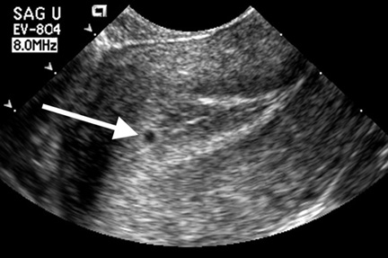
The first sonographic sign that someone is pregnant is the visualization of the intradecidual sign. This can be seen as early as 11 days post conception and appears as a "round fluid collection eccentrically located within an echogenic area adjacent to the endometrial cavity. This sign marks the gestational sac. At 7 weeks, the "double decidual sac sign" is seen, which is produced by the decidua capsularis and the decidua parietalis.
The sign that sonographers should look for to confirm an intrauterine pregnancy is the secondary yolk sac, with the gestational sac and double decidual sign accompanying it. This can be seen at about 5 gestational weeks with transvaginal scans and 6-7 weeks transabdominally. This should be the first implication, since without visualization of the secondary yolk sac, it could be an ectopic pregnancy or other pathology.
Re: Sonographic Documentation
Lauren,
That's a great summation of the events! In the video that Lania posted they mention that the intradecidual sign and the double decidual sign may not been seen in ~35% of pregnancies. Radiopedia seems to agree. Although, they seem to think the percent of pregnancies lacking these signs can be even higher.
https://radiopaedia.org/articles/intradecidual-sac-sign-1?lang=us
https://radiopaedia.org/articles/double-decidual-sac-sign-1?lang=us
Sarah
Re: Sonographic Documentation
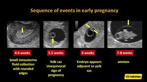
The earliest sonographic sign of of pregnancy is a small intrauterine anechoic, fluid collection with rounded edges which is the gestational sac at around 4-5 weeks. The next sign is a yolk sac appearing within the gestational sac which is an unequivocal sign of pregnancy that occurs around 5.5 weeks. At about 6 weeks the embryo appears adjacent to the yolk sac. These are the first early sonographic signs of pregnancy. Also the double decimal sac sign should be seen at around 5 weeks. The outer ring is the decidua and the inner ring is the chorion. Below I attached a YouTube video that really helped walk me through the steps of what an early normal and abnormal pregnancy appear as sonographically. SUPER HELPFUL!
https://www.youtube.com/watch?app=desktop&v=raIclmdJOPo
Re: Sonographic Documentation
Lania,
This video is amazing!
I'm a dork. I thought the gestational sac would show a yolk sac the moment it became visible. These little details are extremely important and it's imperative that we understand each nuance. Additionally, I like to note that when the embryo is seen at ~6 weeks the heart flicker may also be seen.
To simplify:
Uterine fluid collection with intradecidual sign/double decidual sign (these may not been seen in some IUPs) => yolk sac => embryo (maybe with visible heart flicker => amnion
I don't understand what they mean by "collapsed endometrium" though.
Sarah
Re: Sonographic Documentation
Hey Moderator Buddy!
We learned that the earliest sonographic finding in the first trimester is the gestational sac at around 4 weeks. At this point the uterus should look nice and plump with a small sac that appears cystic adjacent to the fundus. There should be nothing in the sac at this point and would not even be called a gestational sac.
The next finding that would be seen in the first trimester is the yolk sac which is another round echogenic structure with a sonolucent center. The yolk sac would be see around the 5-6 week and should be connected the the gestational sac.
Another sonographic sign that might be seen simultaneously with the other structures is decidualiztion. From my understanding this is the maternals contribution and is made up of the endometrium basal layer.
I believe it is reliable to diagnose a patient when the gestational sac is seen with a properly attached yolk sac. To also answer some of your questions Hailey, I think it is important not to mistaken the GS for a cyst in the endometrium or to mistaken it for fluid in the uterine cavity. I was wondering if you were unsure if a sonographic finding was an abnormality or a pregnancy how would you know if you couldn’t put color(in case it was a baby)? Might be a stupid question ..
Re: Sonographic Documentation
Hi Candee! Please correct me if I'm wrong, but I believe you are mentioning the double-decidual sac sign. It involves the Decidua Capsularis and Parietalis. I thought did a good job describing the decidual endometrium components with drawings and images. For decidualized endometrium, there are three components. The Decidua Basalis is where the blastocyst burrows in and will become the maternal component of the placenta. The Decidua Capsularis encapsulates the remainder of the blastocyst that is not covered by the decidua basalis. The Decidua Parietalis is the remaining area of decidualized endometrial tissue.
To answer your smart question about what to do if you can't put color on a suspected abnormality during potential early stages of pregnancy, my best guess would be to book a follow up appointment on a later date to see if a pregnancy develops or if something else is going on. The hCG blood serum levels should likely to monitored also for changes. I would love to hear someone else weigh in on this if they know more!
Re: Sonographic Documentation
The earliest definitive sign that a sonographer may see of a pregnancy would be in the yolk sac within the gestational sac. This, for me, would be the most definitive sign of an uterine pregnancy because an empty gestational sac could be fluid, a cyst, or a pseudosac. I make the distinction of uterine pregnancy because pseudosacs are created by the bleeding of decidualized endometrium from an ectopic implantation. A yolk sac would be seen at a little over 5 weeks gestational age, whereas the gestational sac could be detected earlier.
Here is a great picture comparing the both:
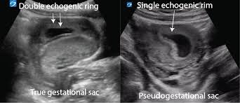
Here is an article on pseudosacs for anyone interested:
https://www.wjgnet.com/2218-6220/full/v4/i3/58.htm#:~:text=A%20%E2%80%9Cpseudosac%E2%80%9D%20is%20a%20collection,pregnancy%20implantation%20(Figure%202).
Re: Sonographic Documentation
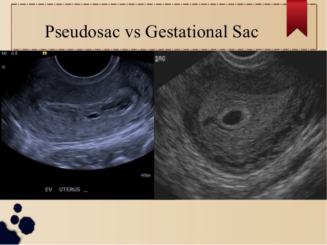
Hey Karen! Your picture confused me a bit. I thought a true gestational sac would be adjacent to the endometrium and not inside of the endometrium like in the picture. The psuedogestational sac seemed more fitting of the true gestational sac. Please let me know.
Re: Sonographic Documentation
Hi Candee,
You're right, maybe that was not the best pic! I think the uterus on the right is flexed in an odd position, making it hard to discern true location. Here may be a better one.
Re: Sonographic Documentation
Oooh I like those ones! The psuedosac definitely looks more distinguishable in that picture. Have you gotten to see a psuedosac yet??
Re: Sonographic Documentation
I am hoping the gestational sac I saw today was not a pseudosac! Patient was 5wk2d pregnant and no yolk sac yet. After this DB I am really wondering...
Re: Sonographic Documentation
Hey Karen!
I believe the yolk sac can appear some around 5-5.5 weeks, so hopefully it will "sprout" so to speak within the next few days. I am curious what the protocol would be going forward for this patient since she is still technically within the time frame. I am wondering if they will send her back for a 6 week scan, although that unfortunately exposes the embryo to more potential energy if it is a viable pregnancy.
Re: Sonographic Documentation
Hey Ms. Candee,
I can definitely understand what you mean. Correct me if I am wrong, but I believe the true gestational sac is outside the endometrial cavity. I remember reading the endometrial cavity would appear as a thin hyperechoic line. I think that is symbolized by the hyperechoic line posterior to the sac.
Re: Sonographic Documentation
Hey Ms. Karen,
Thank you for sharing this. It is an important reminder for us to be thorough as sonographers and not get too comfortable. If someone is moving fast they could easily mistake the two. I like how included the yolk would be a more definitive sign as well.
Re: Sonographic Documentation
Hi Karen! Thanks for sharing that great article. I just bookmarked it for future reference.
In regards to pseudogestational sacs, this website also says that it can be differentiated from an IUP because it is usually oval shaped, is located centrally in the uterus and will not have a thick, echogenic chorionic ring surrounding it.
Re: Sonographic Documentation
Howdy everyone,
The earliest sign of pregnancy on sonogram is a small anechoic structure within the uterus (gestational sac). Even if there is an ectopic pregnancy a round anechoic area may exist in the uterus. In order to confirm an IUP (and rule out pathologies) we must see the yolk sac! It's important to remember that just because there is an anechoic structure in the uterus doesn't mean it's always a pregnancy. We have to be diligent while interrogating other areas as well.
This is an excellent overview of early pregnancy.
https://radiologykey.com/ultrasound-of-the-normal-and-failed-first-trimester-pregnancy/
Sarah
Re: Sonographic Documentation
Hi Sarah, I looked at the resource you shared and a lot of them look like intrauterine cysts.
I liked this 3D representation of an ectopic pregnancy for first trimester. It is a short video with nice visuals!
https://www.youtube.com/watch?v=qtaBidjTThU
Re: Sonographic Documentation
Hi Sarah! The queen of sonographic resources. Thank you for confirming that the gestational sac can also be mistaken as an ectopic pregnancy. I was going on a whim when I explained that on my discussion post as well. No one has corrected me yet...or even responded )-: So I'm happy to come across yours! The yolk sac would be present by the 5.5 week?
I was discussing this with. my CI and he was telling me that the medical field evolved so much, lab test are the best away to confirm anything. From your lab urine to fine the presence of hCG to blood test to find out the gender of the growing baby (at such an early date too)! This quarantine babies are no joke. So excited to see and hear from your clinical journey on Friday!
Re: Sonographic Documentation
Hi Sarah,
I always enjoy your responses you keep also make your point in an understanding form. Some time too much detail can be super confusing. In my response I also included the decidual signs as another "tell-tell" sign of pregnancy. Here a slide show I found on early pregnancy. It does go in to pathology however, which we haven't discussed.
https://www.slideshare.net/walidahmed1276/early-pregnancy-ultrasound
Re: Sonographic Documentation
Great post Sarah! Today we had a first trimester OB patient 6 weeks along and was referred by her OBGYN because they suspected ectopic pregnancy. Sure enough there was a gestational sac visualized in the uterus but no fetal pole or yolk sac. There was a corpus luteum cyst within her ovary with the “ring of fire” sign visualized So HCG was definitely rising. Unfortunately the radiologist had us send her back upstairs to her OB for results and next steps of care. She had an ectopic pregnancy.
I appreciate the resource you posted. I’ll post a photo of the ring of Fire sign visualized around the corpus luteum cyst. One of the sonographers showed me so now I won’t forget it.
I agree to never assume pregnancy just by visualizing the gestational sac. It’s important to sweep thorough thoroughly and document everything seen and not seen.
great job!
-Molly
Re: Sonographic Documentation
Hey Molly!
It is so exciting to hear stories from everyone's clinical experiences, I feel like I am living vicariously through everyone! So sad to hear that your patient experienced an ectopic pregnancy, I can only imagine the helpless feeling everyone in the exam must of felt for her. This is a huge reminder on the importance of understanding embryology and its timeline. Had the patient come in maybe a week or so sooner just visualizing the gestational sac alone would not have been worrisome, especially in conjunction with her hCG levels and corpus luteum cyst! One thing I am curious to learn about is the next steps following an ectopic. Does the body simply dissolve it like the vanishing twin phenomenom or is there medical intervention? Thanks again for sharing your first hand experience!
Re: Sonographic Documentation
Hi Brittany and Chrishawna!
Thank you for your questions and support! I am so eager for you guys to also start clinicals too!!
My CI nor the radiologist stated the location of the ectopic pregnancy. My CI explained how it had all the signs leading to an ectopic and the radiologist deemed it one and sent her upstairs. I would love to know the next steps of care she'll be receiving. One of the other sonographer's said with ectopic cases they can sometimes provide medication to the patient to abort the pregnancy or surgery could be warranted if not. That is very interesting Chrishawna to consider if the ectopic pregnancy could vanish like the "vanishing twin." I suppose maybe that could possibly occur if there was no viability, but with viability a clinical action would need to occur. Super sad! But on the other hand there were a couple of other OB patient's in their first trimesters this week and the heart beats were visualized. That was super exciting to see and somewhat outweighs the bad.
Re: Sonographic Documentation
Hi Molly!!
I'm so excited that you finally got to start clinicals! This seems like a really interesting yet sad case to see.
I'm curious - were you able to see where the ectopic pregnancy was located?
Re: Sonographic Documentation
Hi Molly,
That is so great that you were able to see an ectopic pregnancy so now you know what to look out for. Thank you for sharing as well. I agree as sonographers we have a huge responsibility to check all the areas for any abnormalities. At least we have the knowledge to not just assume by seeing a gestational sac but to not call it that unless we see a yolk sac.
Re: Sonographic Documentation

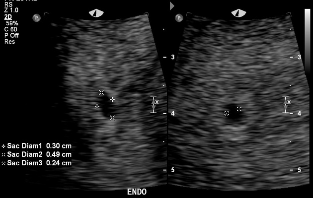
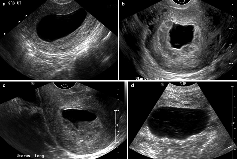
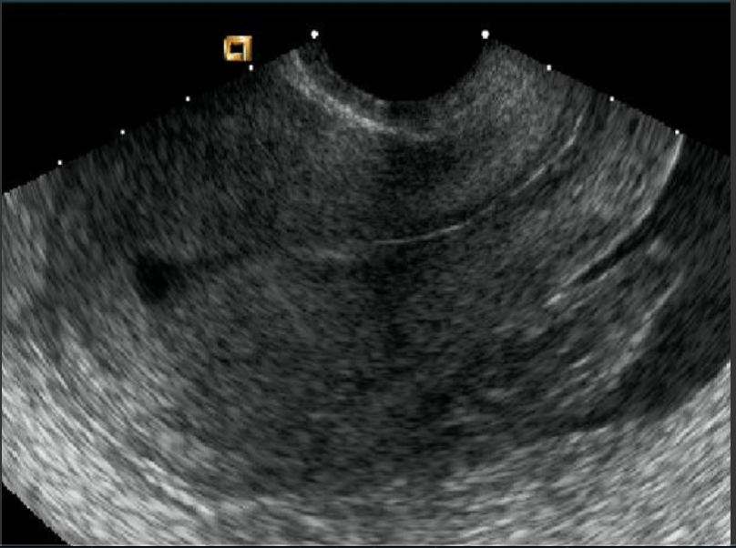
Since we don't really see many measurements in class I thought this is a good representation of how to measure early OB first trimester. I know everyone has a different way of measuring but if this is not the ideal way please lmk
Re: Sonographic Documentation
Hey Ms. Candee,
Thank you for these measurements. These are indeed helpful. Not only does it provide what the images should look like but it also shows how to properly measures them. Awesome! Measurements are so important to track the development of the fetus. I honestly need to go over the proper measurements. All I've got so far is when the gestational sac in 12mm, the yolk sac should have been visualized by this point.
Here's a slideshow I found online regarding intrauterine pregnancy:
https://www.slideshare.net/walidahmed1276/early-pregnancy-ultrasound
Re: Sonographic Documentation
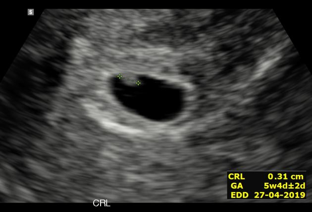
Thanks for sharing! I found this website that showed images of how to measure the MSD and CRL. It is consistent with what you posted.
Just curious, what is image D? is looks like it could a GS with a yolk sac, but the internal echoes seem to be more echogenic than normal. Perhaps it's just a gain issue?
Re: Sonographic Documentation
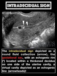
According to our textbook for the class, a sign of a conceptus can be seen transvaginally as early as 3w4d GA when implantation is complete and the blastocyst is fully implanted in the decidua. It will look like a small, fluid collection with an echogenic border just off center of the endometrial cavity and is called the intradecidual sign. At this point, the chorionic cavity (aka gestational sac) is seen and is filled with chorionic fluid, giving it a cystic-like appearance.
However, as Dr. Wilson was explaining in lecture, it is safer to definitively call a IUP when the secondary yolk sac is seen within the gestational sac, usually around 5-5.5 weeks GA. The secondary yolk sac (aka umbilical vesicle) is responsible for providing nourishment to the embryo and also hematopoiesis. Assuming the patient is being examined partially due to detectable hCG blood serum levels, we would suspect the patient is pregnant. At this early stage, we want to make sure to rule out an ectopic pregnancy as there are, to my knowledge, poor outcomes for ectopic pregnancies. Seeing the secondary yolk sac inside the gestational sac would prove it is an IUP and not ectopic.
Here is a YouTube Video that walks through the sonographic signs of early pregnancy.
Re: Sonographic Documentation
2. As early as 3 weeks and 4 days gestational age, the chorionic sac can be visualized using ultrasound. The chorionic sac, AKA gestational sac, is the first sonographic sign of intrauterine pregnancy. In ULS the term used for this image is intradecidual sign:
IMAGE 1 HERE ("intra")
It is important to note that this location is not within the endometrial cavity.
IMAGE 2 HERE ("Sequence")
Between 5-6 weeks, the embryo adjacent to the yolk sac can be identified transvaginally. This is a good sign in terms of the prognosis of pregnancy because failure to identify the yolk sac once the gestational sac has reach 12mm suggest failed pregnancy. The location of embryo being in the uterus is important as well to rule out ectopic pregnancy. It is crucial to measure the yolk sac at this stage as well. When the yolk sac is greater than 6mm at the 5th week is highlights an abnormal pregnancy. Presence of cardiac activity is important at this time as well; normally measuring greater than 5mm with an average of 110 BPM. By 7 weeks the double decidual sign will be present. These criteria combined can confirm an intrauterine pregnancy.
Re: Sonographic Documentation
Hi Nailiah!
I love the visuals you provided for the stages of a developing Intrauterine pregnancy. It’s very helpful! Paying close attention to dates, measurements, and which structures are present or absent will be super important in our success. Those all definitely confirm pregnancy also the corpus luteum cyst is a good indicator.
Clinical’s are sure confirming just how much pressure we have to provide good diagnosable exams for patients and how intense/challenging some exams can be.
good job!
-Molly